User:Courtney Krawczyk/Notebook/Biology 210 at AU: Difference between revisions
No edit summary |
No edit summary |
||
| Line 1: | Line 1: | ||
'''Observing and Identifying Algae and Protists''' January 29th, 2015 | '''Observing and Identifying Algae and Protists''' January 29th, 2015 | ||
'''Purpose''' The purpose of this study was to | '''Purpose''' The purpose of this study was to observe and identify different organisms from the culture we had made in the previous lab. The second part of this lab consisted of plating serial dilutions of the mixture of bacteria species onto nutrient agar petri dishes to observe at a later time. | ||
'''Materials and Method''' First, I took a sample for one area of the hay infusion culture (top right side) and made a wet mount slide. Using the microscope I looked at the slide in search of any organism, then observed and recorded data on what I saw. I then repeated this step, but this time I took a sample from a different part of the hay infusion culture (the bottom left of the jar). After observing and recording this information, I used a dichotomous key to help identify what organisms I may have seen that were present in the culture. The second part of the experiment was setting up agar plates to allow bacteria to divide many times to form colonies and look at on another date. First four nutrient agar plates and four nutrient agar plates plus tetracycline were obtained and labeled either 10^-3, 10^-5, 10^-7, and 10^-9 (each type of plate was labeled with a different dilution). After labeling the plates, the hay infusion culture was swirled and mixed thoroughly. Four different tubes full of 10mLs sterile broth each, were also labeled 10^-2, 10^-4, 10^-6, and 10^-8. Then 100 microliters of the culture was assed to the 10mls of broth in the 10^-2 tube. THen 100 microliters of broth from that tube was pipetted into the 10^-4 tube (again to the 10^-6 and finally from that tube to the 10^-8 tube). After the dilutions were completed, 100 microliters from the 10^-2 plate was pipetted on the the 10^-3 nutrient agar plate and another 100 microliters onto the 10^-3 nutrient agar plate plus tetracycline plate. This was repeated for each tube and agar plate, the 10^-4 dilution on the 10^-5 plates ,the 10^-6 on the 10^-7 plates, and the 10^-8 on the 10^-9 plates. The plates were then placed agar side up on a rack and left to incubate for one week. | '''Materials and Method''' First, I took a sample for one area of the hay infusion culture (top right side) and made a wet mount slide. Using the microscope I looked at the slide in search of any organism, then observed and recorded data on what I saw. I then repeated this step, but this time I took a sample from a different part of the hay infusion culture (the bottom left of the jar). After observing and recording this information, I used a dichotomous key to help identify what organisms I may have seen that were present in the culture. The second part of the experiment was setting up agar plates to allow bacteria to divide many times to form colonies and look at on another date. First four nutrient agar plates and four nutrient agar plates plus tetracycline were obtained and labeled either 10^-3, 10^-5, 10^-7, and 10^-9 (each type of plate was labeled with a different dilution). After labeling the plates, the hay infusion culture was swirled and mixed thoroughly. Four different tubes full of 10mLs sterile broth each, were also labeled 10^-2, 10^-4, 10^-6, and 10^-8. Then 100 microliters of the culture was assed to the 10mls of broth in the 10^-2 tube. THen 100 microliters of broth from that tube was pipetted into the 10^-4 tube (again to the 10^-6 and finally from that tube to the 10^-8 tube). After the dilutions were completed, 100 microliters from the 10^-2 plate was pipetted on the the 10^-3 nutrient agar plate and another 100 microliters onto the 10^-3 nutrient agar plate plus tetracycline plate. This was repeated for each tube and agar plate, the 10^-4 dilution on the 10^-5 plates ,the 10^-6 on the 10^-7 plates, and the 10^-8 on the 10^-9 plates. The plates were then placed agar side up on a rack and left to incubate for one week. | ||
'''Data and Observations''' After viewing the hay infusion culture, it was clear that some brownish moldy looking type substance was floating on the top of the water in the jar. The liquid mixture appeared to be completely brown. It also had a strong moldy smell. Below is a picture of what the hay infusion culture looked like. When looking at the slide that was taken from the top left of the culture, I found many moving organisms that appeared to possibly be peranema. There were up to ten in view at one time. the were clear in color with a little green internal coloring. The cell was elongated and slightly rounded. When viewed in the 40x | '''Data and Observations''' After viewing the hay infusion culture, it was clear that some brownish moldy looking type substance was floating on the top of the water in the jar. The liquid mixture appeared to be completely brown. It also had a strong moldy smell. Below is a picture of what the hay infusion culture looked like. When looking at the slide that was taken from the top left of the culture, I found many moving organisms that appeared to possibly be peranema. There were up to ten in view at one time. the were clear in color with a little green internal coloring. The cell was elongated and slightly rounded. When viewed in the 40x magnifications, the organisms appeared to be slightly vibrating. I also saw a cluster of what looked like tiny green cells. The individual cells were round but formed an irregular shape when linked together. This may have been gonium. The sample taken from the bottom right of the jar also appeared to have life present when viewed under the microscope. First I saw an organism that appeared to me moving very quickly. It was clear with some green internal coloring and very circular. The next organism I observed was clear with light pink coloring inside. It was very motile and I think this may have been Blepharisma. I estimate that I saw at least 150 of these organisms. Last I saw what I believe to be gonium again. They were very small green clusters of cells. The motile organisms meet the needs of life because they are made of at least one cell, they needed the jar to remain open for air, and they also needed food to survive. If the hay infusion culture continued to "grow" for another two months, I predict that the smell would definitely become much more pungent. I also think that much more mold would be visible to the naked eye growing within the culture. | ||
[[Image:biolab2.jpeg]] | [[Image:biolab2.jpeg]] | ||
Revision as of 18:47, 29 January 2015
Observing and Identifying Algae and Protists January 29th, 2015
Purpose The purpose of this study was to observe and identify different organisms from the culture we had made in the previous lab. The second part of this lab consisted of plating serial dilutions of the mixture of bacteria species onto nutrient agar petri dishes to observe at a later time.
Materials and Method First, I took a sample for one area of the hay infusion culture (top right side) and made a wet mount slide. Using the microscope I looked at the slide in search of any organism, then observed and recorded data on what I saw. I then repeated this step, but this time I took a sample from a different part of the hay infusion culture (the bottom left of the jar). After observing and recording this information, I used a dichotomous key to help identify what organisms I may have seen that were present in the culture. The second part of the experiment was setting up agar plates to allow bacteria to divide many times to form colonies and look at on another date. First four nutrient agar plates and four nutrient agar plates plus tetracycline were obtained and labeled either 10^-3, 10^-5, 10^-7, and 10^-9 (each type of plate was labeled with a different dilution). After labeling the plates, the hay infusion culture was swirled and mixed thoroughly. Four different tubes full of 10mLs sterile broth each, were also labeled 10^-2, 10^-4, 10^-6, and 10^-8. Then 100 microliters of the culture was assed to the 10mls of broth in the 10^-2 tube. THen 100 microliters of broth from that tube was pipetted into the 10^-4 tube (again to the 10^-6 and finally from that tube to the 10^-8 tube). After the dilutions were completed, 100 microliters from the 10^-2 plate was pipetted on the the 10^-3 nutrient agar plate and another 100 microliters onto the 10^-3 nutrient agar plate plus tetracycline plate. This was repeated for each tube and agar plate, the 10^-4 dilution on the 10^-5 plates ,the 10^-6 on the 10^-7 plates, and the 10^-8 on the 10^-9 plates. The plates were then placed agar side up on a rack and left to incubate for one week.
Data and Observations After viewing the hay infusion culture, it was clear that some brownish moldy looking type substance was floating on the top of the water in the jar. The liquid mixture appeared to be completely brown. It also had a strong moldy smell. Below is a picture of what the hay infusion culture looked like. When looking at the slide that was taken from the top left of the culture, I found many moving organisms that appeared to possibly be peranema. There were up to ten in view at one time. the were clear in color with a little green internal coloring. The cell was elongated and slightly rounded. When viewed in the 40x magnifications, the organisms appeared to be slightly vibrating. I also saw a cluster of what looked like tiny green cells. The individual cells were round but formed an irregular shape when linked together. This may have been gonium. The sample taken from the bottom right of the jar also appeared to have life present when viewed under the microscope. First I saw an organism that appeared to me moving very quickly. It was clear with some green internal coloring and very circular. The next organism I observed was clear with light pink coloring inside. It was very motile and I think this may have been Blepharisma. I estimate that I saw at least 150 of these organisms. Last I saw what I believe to be gonium again. They were very small green clusters of cells. The motile organisms meet the needs of life because they are made of at least one cell, they needed the jar to remain open for air, and they also needed food to survive. If the hay infusion culture continued to "grow" for another two months, I predict that the smell would definitely become much more pungent. I also think that much more mold would be visible to the naked eye growing within the culture.


Conclusions and Future Directions I predict that there will be much more growth of bacteria in the nutrient agar plates than the plates with the tetracycline antibiotic. The least diluted agar plates will probably have the most visible bacteria colonies, while the most diluted plates will probably show much less growth. It is also possible that fungus may appear in the agar plates as well.
CK
Study of Biological Life in an Ecosystem January 28, 2015
Purpose In this study both biotic and abiotic components were observed in an ecosystem. This portion of the study includes analyzing the specific 20 x 20 meter transect, specifically looking at what makes up this ecosystem. After a collection of the biotic and abiotic components was taken, a hay infusion culture was created to later observe protists, bacteria, and changes in the culture overtime.
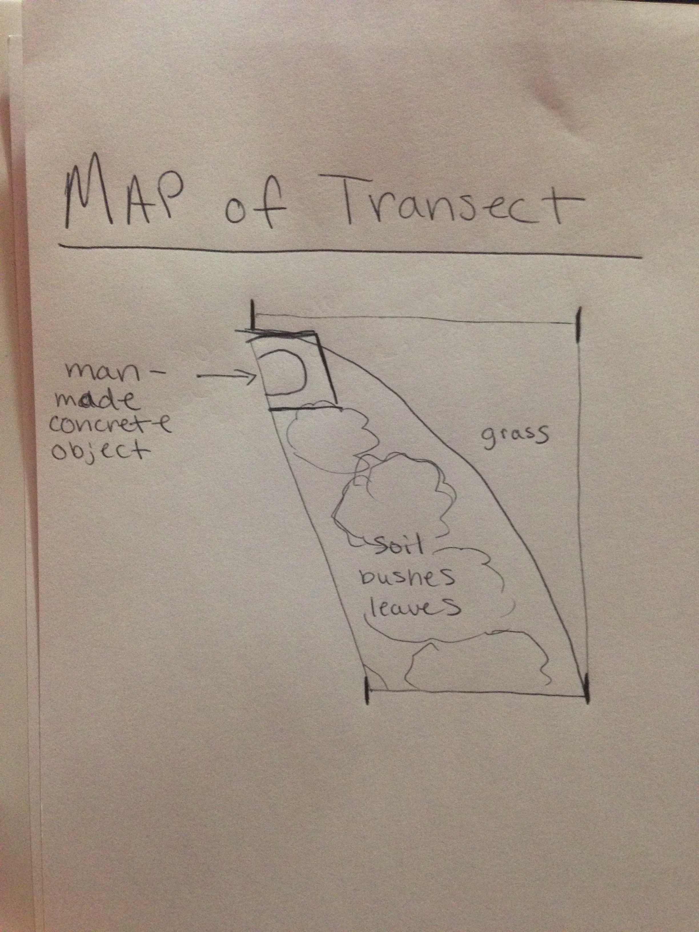
Materials and Method After being assigned a specific transect to observe, my lab partners and I first drew a map of the area. Everything was labeled that was visible, for example, plants, soil, rocks, leaves, and even manmade objects. A list of five biotic and five abiotic components found in the transect was also composed. Then a sample consisting of soil and plants was put into a 50mL conical tube. After returning back to the lab, 11.7 grams of the sample was placed into a jar with 500mLs of water. Then 0.1 gm of dried milk was also added to the sample and mixed for about 10 seconds. The jar, known as a hay infusion culture, was then labeled with our transect number (5), and left in the lab for a week with the lid off.
Data and Observations Attached below is a hand drawn map of the transect as well as other pictures of the area. The 20x20 meter transect consisted of a larger grass area. It also contained an area of soil with bushes, weeds, berries, dead leaves, and twigs. Coming into one corner of our transect was also a man made concrete sign. 5 biotic components of the transect -bushes (possible rose plants) -grass clumps under bushes -grass -berries -weeds 5 abiotic components of the transect -soil -dead leaves -snow -manmade stone structure -wood chips/twigs
Conclusion and Future Directions It can be concluded that both biotic and abiotic components make up the transect. I predict that next week when observing the hay infusion culture, it will probably appear moldy and have a noticeable smell. When observing slides with samples taken from the culture, I expect to see protists within the sample.
http://openwetware.org/images/2/28/Map_for_bio.jpeg
CK
January 21, 2015
This is my first wetware post
CK