User:Aurriel Fenison/Notebook/Biology 210 at AU
March 20, 2014
Embryology Lab
Objective: The objectives for this lab was to learn the stages of embryonic development and then compare embryonic development in different organisms. One of main objectives of this lab was to set up an experiment to study how environmental conditions affect embryonic development.
Procedures:
Read and analyze a published paper about the effect darkness has on zebrafish development, make a hypothesis, prediction, and experimental plan. Then set up the conrol group and the test group in covered petri dishes. Each group tests one variable so there will be at least two petri dishes of embryos. Use 20 mls of Deerpark water and 20 healthy translucent embryos per dish. Use a dropper to transfer the eggs to the dishes with the appropriate water. Organize the observation schedule and procedure. Make observations and carefully record those over the next two weeks. When embryos are 4-5 days old, removes 10 mls of water with an empty egg cases and add 25 mld of fresh water. Save any dead embryos in paraformaldehyde that will be provided. one week after the experiment has begun, remove 5 mls water with any egg cases and add 5 mls fresh water. Preserve 1-3 embryos from the control and the experimental groups in paraformaldehyde. Between 1 week and 2 weeks incubation time, remove 5 mls and add 10 mls. Fee two drops of paramecium starting after one week. this can be continued just when the water is changed. Make final observations and measurements at two weeks. at that time the surviving embryos will be collected and placed in an aquarium for safe keeping. Prepare tables and graphs and any calculations for your presentation.
Raw data: All of the graphs can be found here: https://drive.google.com/file/d/0B7FiMC1bByX9aDNzdkxTMzRKTExvQmppNzl5QnhDVmpqSnVj/edit?usp=sharing
Conclusion:
One of the main conclusions that were found was the experimental group swam a lot slower than the control group and had an average heart rate that was faster compared to the control. Other than this, the experimental zebrafish's development wasn't much different from the control. The control zebrafish on average had a larger body by about 100 micrometers, but as far as the other aspects of development keeping the experimental zebrafish in the dark did not effect the development of the zebrafish significantly different. In every aspect, the control group was slightly more developed, but the experimental groupd was not far behind. If the experimental group was in the wild, they would still be able to survive. However, one thing that was noted was that the experimental zebrafish had more zebrafish dead by day 11 than the control zebrafish. Unfortunately, by day 14 some unknown factor killed all of the control zebrafish so all of the observations from the both sets of zebrafish were made from fixed slides at day all. This lab was very interesting and fun to do, and besides the unknown factor with the control zebrafish dying, there wouldn't be anything that I would change about this experiment.
Aurriel Fenison
February 26, 2014
Lab 6 Objective:
To determine species observed in the hay infusion
Procedures:
see lab 2&3
Raw Data:
link to table: https://docs.google.com/spreadsheet/ccc?key=0ArFiMC1bByX9dGFLNWltc1A3R1BJNTlwMGNkbGJkNEE&usp=sharing
Conclusion:
For our first bacteria, the Aeromonas sp was gram-negative but we got that the bacteria was gram-positive. Besides that everything else that we observed both bacteria to have, the information we received on them was the same. It was unclear however if the Aeromonas sp. was mobile or immobile.
Aurriel Fenison
February 13, 2014
Lab 5 Objective:
To understand the importance of invertebrates and to learn how simple specialized cells to overall body parts have become more complex.
Procedures:
Describe the movements of the three different types of worms and how they movement relates to their body structure. Then randomly select five organisms in the petri dish and identify each of them as close as you ca. If your transect does not have five organisms, then use the organisms in the West Virginia petri dishes. Use the dissecting microscope to view the organisms, and then fill out the table below. Note the size each organisms is and the range they fall in. Which organisms is the largest? Which is the smallest? What kids of organism are the most common in the leaf litter? After the organisms from the transect have been examined,look at the groups of vertebrates as they are organized in the textbook and identify five who might inhabit the transect. At least two must be bird species. Determine the classification of each, starting with the phylum through the species. Then describe what biotic (possibly food) and abiotic characteristics of the transect would benefit each species. As a final summary figure, construct a food web based on the organisms you have observed in your transect, or from West Virginia. Carefully read pages 1150-1151 of the Freeman and model your diagram after Figure 56.4.
Raw data:
The first worm that we examined was the Platyhelminthes, flat worm, which are considered acoelomates. These worms were fairly wide in diameter, they moved slow and in order for them to move they go back and forth and slowly inch where they need to go. The second warm that we looked at was Nematodes, round worms, and specifically the Cephalobus Genus. These worms were long and thin and circular. Depending on how long their bodies are, sometimes they can be found curled up. These worms move about by wiggling side to side. The last worm that we looked at was the Annelida coelomate, or the earth worm. This worm has a long body as well with tons of rings around them. On either side of the worm has pointy ends, and they use accordion-like movement. As we were looking though the Berlese Funnel, we only found one organism so we classified the other 4 from West Virginia. In the link, there is table with the kind of organism and a brief description of the organism and their relative size. The range in size for the organisms were from .2 - 200 mm and the smallest was the soil mite and the largest was the mosquito. We only found one organism in our transect, so it is unclear which organisms are most common to in the leaf litter. The food that the organism in my transect and the organisms from West Virginia would eat would be leaves, dead organic matter as well as other organisms such as other spiders or other smaller insects. Some of the abiotic characteristics that the organisms would benefit from is the soil. They wouldn't really benefit from the concrete because it is mostly occupied by humans and there is no advantage that these organisms can use from the concrete.
The one organism that we found in our transect was the soil mite and it can be classified as the following: Kingdom: Animalia Phylum: Arthropoda Subphylum: Chelicerata Class: Arachnida Subclass: Acari Superorder: Acariformes Order: Oribatida
The two Springtails can be classified in the following: Kingdom: Animalia Phylum: Arthropoda Subphylum: Hexapoda Class: Entognatha Subclass: Collembola
The centipede can be classified as the following: Kingdom: Animalia Phylum: Arthropoda Subphylum: Myriapoda Class: Myriapoda
The mosquito can be classified as the following: Kingdom: Animalia Phylum: Arthropoda Class: Insecta Order: Diptera Suborder: Nematocera Infraorder: Culicomorpha Superfamily: Culicoidea Family: Culicidae
here is an example of a food web or what might possibly inhabit my transect: https://docs.google.com/spreadsheet/ccc?key=0ArFiMC1bByX9dHlqbmUyanlVaXpHQktrdG5wXy1KVFE&usp=sharing

Conclusion:
At the end of this lab, we learned how to identify the three different type of worms as well as examine and describe the invertebrates that we found in our transect. I feel like this lab was very well pre[ared because for those of us who did not have 5 invertebrates, some invertebrates were provided for us. One thing that was challenging about this lab was trying to measure the length of the organism because the dissecting scope doesn't have any thing to take measurements with so we were told just to guess or look it up. Another thing that made this lab a little difficult was constructing the food web with animals from our transect. A reason for this is because most of the organisms that we studied were microscopic and mostly invertebrates and the book was not very helpful for the criteria that we needed to construct this food web. One thing I would suggest is in class to go over a food web and some of the hierarchy of it, so when we are constructing our food web we have some idea what actually happens in nature.
Aurriel Fenison
February 12, 2014
Lab 4 Objective:
The objective of this lab is to understand the characteristics and diversity of plants as well as to appreciate the function and importance of Fungi.
Procedures:
For this lab describe the five plants and where they were found. Use Table 1 as a guide to keep all the information organized. Then use the information to identify the major group from the table above and reference additional resources to possibly determine the genera for each of the five samples. Then briefly describe the vascularization in each of the plants from your transact. Briefly describe the shape, size, and cluster arrangement of the leaves from the transect plants. If there are no leaves, examine the attachment site or evidence of leaves in the area. Once that is completed, identify the seeds you brought back from the transect as either monocot of dicot. See if there is evidence of flowers or spores. Take a look at some of the samples with the dissecting microscope and decide if they are fungi and which of the three groups they belong to. Draw a picture of one and describe it in your notebook as well as why you think it is a fungus. Lastly answer the question of What are the Fungi sporangia and why they are important?
Raw Data:
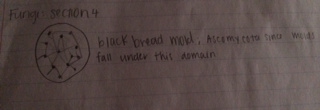
The plants that we retrieved from our transect a tree leaf that was located near the edge of the soil near the concrete by the bench, then we gathered another leaf off of the ground from the rose bush with what two seed-like pods. We also collected some grass from the edge of the transect where the concrete, dirt and grass meet. Around this area as well we grabbed a two leaf clover and on the concrete near the bench was some moss. Table 1 explains the location, description (shape and size), vascularization, the different type of leaves and special characteristics and the seeds, evidence of flowers or other reproductive parts. Four out of the five plants that we collected had Xylem and Phloem for their vascularization system. The leaf from the Black Oak, Rush Bush, and the clover all had branched leaf veins while the grass had a parallel veins. The only plant that had a seed was the the leaf from the rose bush and it was a monocot. While we were studying Fungi, we discovered that the sporangia is when the hype grows upward and they form small, black, globelike structures. The sporangia is important because it contains the spores that Fungus release for reproduction. The Fungus that we looked at under the dissecting scope was black bread mold from the Ascomycota domain. Below is a picture of of how the black bread mold looked.
Conclusion:
In Conclusion, we were able to identify the different types of plants, as well as their vascularization, special characteristic, seeds and evidence of flowers or other reproductive parts. We also looked at Fungus and had to identify it. The fungus that my group looked at was Black Bread mold that is commonly found on foods. One thing that would have made this lab a little better was if we went over in detail exactly what the different sections of Table 1 meant and how detailed they should have been. For example there is a section label "vascularization", but it is unclear whether we were just supposed to state whether or not the plant was vascular or not or what type of vascularization was used. Other than this small detail, the lab gave us a lot of background information about the plants and fungus and their different parts.
Aurriel Fenison
2/6/14, lab 1 data
Good job on your write up! Work on creating a map of your transect so we can get a detailed image of your land and know where you samples came from! We will talk more about this Wednesday.
AP
February 5, 2014 Lab 3
Objective:
To understand the different characteristics of bacteria as well as to understand how DNA sequences are used to identify different species. Another objective of this lab os to observe antibiotic resistance and how it can affect the growth of bacteria that grows with antibiotics and those that do not.
Procedures:
For this lab explain why the appearance or smell might change week to week. Then when observing whether the different types of bacteria are antibiotic resistant or not, observe where there is any differences I'm the colony types between the plates with vs without antibiotic. What does this indicate? What is the effect of tetracycline on the total number of bacteria and fungi? Then note how many species of bacteria are unaffected by the tetracycline. Find out how tetracycline works and the types of bacteria that are sensitive to this antibiotic. Describe the cells and any type of motility as well as draw each of the organisms that you observe.
Raw Data:
The appearance and smell of the Hay Infusions will change from week to week because the decomposers of the niche will start to decompose the little bit of life that is in the niche. Also most of the time the cap is kept on the bottle and there is not oxygen getting to the organisms so they are using anaerobic respiration which is causing the smell. While observing our Hay Infusions, we noticed that everything was still at the bottom, but the grass and the few roots also sunk to the bottom. Although the water is clearer than last week, but is still a mirky brown. The Hay Infusion still has a distance odor, but it is not as bad as the previous week. After we observed the results of our bacteria in four petri dishes without antibiotic and three petri dishes with tetracycline. The effect that tetracycline has on the bacteria is that it prevents the the attractant of tRna to RNA complex. The common types of bacteria that tetracycline kills are E. Coli, Haemophilus influenza, Mycobacterium tuberculosis, Pseudomonas aeruginosa ("Tetracycline"). What we observed was the plates with antibiotics had a lot less bacteria than the plates without. More specifically there was more variety of bacteria of on the petri dish that was without tetracycline compared to the one that had it.This means that this particular bacteria is not antibiotic resistance because the tetracycline did not kill all the bacteria. However, even though the tetracycline did not eliminate all the bacteria, it greatly reduced the number that was able to grow. One thing that we observed was that the number of bacteria was lower at 10-3 but higher for 10-7 for the bacteria that did not have any antibiotic on the dish. Most of the colonies for both the bacteria with and without antibiotics appeared to be in circular arrangements and irregular. The bacteria consisted of three different colors: orange, white and beige. The nutrient plate had more white colonies out of the three colors. Also unlike the nutrient agar, the nutrient bacteria had an abundance of all three colors while the tetracycline seemed only to produce mostly large orange colonies and a few smaller beige bacteria colonies.The bacteria on the tetracycline plate were a lot bigger compared to the nutrient plate of bacteria and the orange colonies were dominant.This may be because the nutrient plate of bacteria did not have a lot of room to grow because the plate was so overpopulated with not a lot of room to grow. While observing the three different types of bacteria--nutrient white, nutrient orange and tetracycline white-- under the microscope, it was discovered that the nutrient white bacteria were gram positive and were motile with circular dots and said it was a Tetrad. The nutrient orange bacteria were also gram positive, but they were not motile and they were arranged in several clusters and were identified to also be Tetrads. The last type of bacteria was tetracycline white and it was gram negative, it had a linear shape and it not motile. Unlike the nutrient white and orange, the tetracycline white was more spread out and was identified as a Diplobacilli.
Conclusions:
While observing our bacteria that the nutrient plate had more white colonies out of the three colors, which lead us to determine that tetracycline killed most of the white bacteria. Since on the nutrient plates had an abundance of white colonies, when the tetracycline killed of most of those bacteria, it allowed the orange and beige bacteria to grow to closer to their full size because they had a lot less competition for space. The identification for the two nutrient petri dishes were Tetrads and the identification for the tetracycline petri dish was Diplobacilli. A useful tool that would help make this lab a little easier, is to actually have use the Bacteria Colony Morphology in practice as well as the Free-Living Protozoa because they are two completely different worksheets and it would be a lot better to have had some practice with the worksheet that we will actually be using.
Works Cited: "Tetracycline".Chemistry and chemical biology of tetracyclines. Tetracyclines. n.d. Web. 5 Fe. 2014.
Aurriel Fenison
January 26, 2014
Lab 2
Objective:
To underhand how to use a dichotomous key and the characteristics of Algae and Protists, by observing several different types of organisms. Also to observe our Hay Infusions and use the dichotomous key to identify any organisms that might be in our niche.
Procedures:
Observe the culture that is developing in your niche. not the smell, and describe the appearance. Also not whether there is any apparent life on the top of the water such as molds or green shoots. Then take a few samples for microscopic observations. Take samples from two different niches: one from the top and one from the bottom. Note why the organisms may differ near the plant and away from the plant. Why might this be? Carefully use a dropper to place a small drop of liquid from the culture onto a microscope slide and place a cover slip on top. Draw organisms that you observe. Characterize at lease three different organisms from each of the two areas. Are the organisms mobile or immobile, protozoa, algae, or others? are they photosynthesizing or not? See if you can identify any of these with the key. Measure the organisms size on the Ocular micrometer. Then choose one of the organisms and describe how this species meets all the needs of life as described on page 2 in the Freeman text. If the hay infusion coulter had been observed for another two months what changes would you expect to occur? What selective pressures affected the composition of your samples?
Raw Data:
As we were observing our Hay Infusion, we noticed that it had murky water. Also all of the dirt had settled to the bottom along with a few grass strands and one dead leaf. There was a light gray film-like layer on top of the water. The Hay Infusion had a very foul odor and we noted that the one grass strand that was floating on top of the water had some mold on it. Besides the glass, there was no apparent life that is visible to the naked eye.
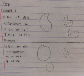
Then we took some samples from the very top of the water near the grass and then right under the grass at the very bottom. Organisms that are near the bottom of the water are most likely protists and costume the nutrients. The ones near the top of the water most likely photosynthesize because they are closer to the top and have easy access to sunlight. The selective pressures that would affect our transect would be the fact that they were in a closed jar. So the pressure of the jar would affect the organism cause it is not the same as if they were out in the environment.
We observed several organisms from both the top of our transect and the bottom. However, all of the organisms that were observed from the top and the bottom niche were colpidium organisms. They were extremely motile and most likely used cilia to move around, they are apart of the protozoa family. They range in size 70-40 micrometers. The ones measured at the top of the transect were from 60-70 micrometers while the organisms measured at the bottom were slightly smaller. They ranged in size of 40-50 micrometers The Colpidium is made up of cells, and in these cells are hereditary or genetic information that can be passed on to their offspring. These organisms use food that they consume to use as energy to survive.


Conclusion: This lab consisted of mostly observing organisms under the microscope and trying to identify them. In our niche we were able to discover the same type of species at the two different habitats from the Dichotomous key, which is what the objective of this lab was. This lab was done very well, and there was no problems and therefore nothing that needs to be changed in future classes.
January 22, 2014
Lab 1
Objective:
To understand the biotic and abiotic characteristics of a niche
Procedures:
Observe a 20x20 ft transect located between 4 popsicle sticks. Then describe the general characters of the transect, such as the location, topography, etc. After observing the niche, list the abiotic and biotic components of the transect. Then use a 50mL conical tube to scoop up a sample and place it in the container. Weigh about 10 to 12 grams of the dirt in a plastic jar with 500 mLs of deep dark water. Then add .1 gm of dried milk and gently mix the mixture. This will be used for the Hay Infusion, which will be observed next lab period.
Raw Data:
Our transect is located in the middle of the quad.The topography of our transect consisted of 1/3 of grass, a rose bush, soil, concrete and stone all at ground level. Also we noticed that there was a bunch of dead leaves on the ground and the rosebush appeared to be dead as well. We were then asked to observe the abiotic and biotic niches for our transect. For the abiotic features, we observed our transect had concrete, stone and soil. For our biotic features, we noted that our transect consisted of rush buses, grass and some small plants which are known as the Native Scrappy Leaf plants. We also pointed out how there are organisms living in the soil. The sample of our niche weighed about 10.9 grams.
Conclusion:
This lab did not consist of any measurements that could be bettered for another lab. Lab 1 consisted mostly of observing and describing the characteristics of our transect.
Aurriel Fenison
assigned user name and lab notebook and entered text successfully
Aurriel Fenison _ yeah! MB


