User:Ambuj Suri/Notebook/Biology 210 at AU: Difference between revisions
Ambuj Suri (talk | contribs) (New page: Test post.) |
Ambuj Suri (talk | contribs) |
||
| (38 intermediate revisions by 2 users not shown) | |||
| Line 1: | Line 1: | ||
Test post. | == Lab 6-8 (2/19/15-3/5/15) == | ||
'''Purpose:''' | |||
The purpose of this experiment was to determine the effects of caffeine on embryonic development, using zebrafish as a model organism. | |||
'''Materials & Methods:''' | |||
Preliminary research was conducted to learn about the possible effects of caffeine on embryonic development of zebrafish. Published papers were used to develop predictions and experimental plan. | |||
Two petri dishes were acquired and labeled as caffeine negative (control) and caffeine positive (experimental). The control environment was prepared by placing 25 mLs of distilled water in a petri dish using a graduated pipette. Additionally, the experimental environment was prepared by placing 25 mLs of 25 mg/L caffeine in the petri dish labeled caffeine positive. 20 healthy zebrafish embryos 24 hours post-fertilization (hpf) were placed in each of the dishes. The embryos were observed under a dissecting microscope to determine developmental stage based on motility, color, and size. | |||
The next day after experimental set-up, dead embryos were removed from each of the petri dishes. In addition, the developmental stage of the living embryos was assessed based on size and physiological features of the embryo. | |||
On the fourth day, 10 mL of solution, debris, and dead fish were removed from each petri dish. To account for removed or evaporated solution, 25 mL of the water and caffeine solutions were added to the control and experimental groups, respectively. The developmental stage of the embryos was recorded. | |||
On the seventh day, the zebrafish were again examined to determine their developmental stage and physiological features. Thereafter 10 mL of caffeine and water were added to account for evaporated water. Additionally, one fish from each of the petri dishes was removed and placed in paraformaldehyde for further examination. | |||
On the eleventh and fourteenth days, debris and dead fish were removed from each petri dish. The approximate developmental stage was recorded along with physical health, such as heart rate, pigmentation, and morphological features. On the fourteenth day, the experiment was terminated by removing dead fish, placing live fish in fresh environments, and discarding the petri dishes. Final observations were recorded and analyzed. | |||
'''Data & Observations:''' | |||
Table 1: Zebrafish Development Over Two Weeks | |||
http://i1382.photobucket.com/albums/ah252/ambujs/f20ca1bb-7e31-4927-af1c-00111f0e3cab_zps7vllcrjl.png | |||
Legend: Table 1 illustrates the number of zebrafish embryos and hatchings that were dead and alive per observation. It also shows the color, motility and average stage of development of the zebrafish in each group. The zebrafish in the caffeine positive environment were consistently underdeveloped than those in the caffeine negative environment. Additionally, the survival rate of the zebrafish in the caffeine environment is lower than that of the zebrafish in the control environment. | |||
'''Conclusion''' | |||
The zebrafish in the experimental group had decreased motility, greater pigmentation, shorter body length, shorter body length, and slowed developmental timelines. In addtion, the number of living zebrafish in the caffeine rich environment was five fish lower than those in the control. Based on these results, it is concluded that caffeine negatively affects embryogenesis in zebrafish relative to the control. | |||
== Lab 7 (2/26/15) == | |||
'''Purpose:''' | |||
The purpose of this experiment is to use DNA sequences to identify species of bacteria found in the transect using polymerase chain reaction (PCR), gel electrophoresis and DNA sequencing to distinguish the 16S gene, which is highly variable. | |||
'''Materials & Methods:''' | |||
Samples from a hay infusion were diluted and plated onto nutrient agar and tetracycline treated plates. Bacteria were left to grow on the plates over the next week. Two colonies from each environment were isolated and transferred to 100 microliters of water in four 200 microliter tubes. | |||
The four tubes were incubated at 100 degrees celcius in a heated water bath. After ten minutes, the samples were centrifuged ay 13,400 rpm for 5 minutes. Following centrifugation, 5 microliters of the resulting supernatant fluid was placed with 20 microliters of PCR mixture (including primers and polymerase) in a new labeled tube. | |||
After one week, the PCR product was stained and placed in an agarose gel in a gel electrophoresis apparatus. Using a P-100, the samples were pipetted into wells on the gel along with a ladder. The gel was placed with the well facing the negative charge so that the DNA product would travel to the positive end after initiating the electrical field. | |||
Two of the remaining samples of the PCR product from the tetracycline-rich plates were sent for sequencing. The DNA sequences were then copied and pasted into a Blast software to determine the genetic identity of the bacterial samples. | |||
'''Data & Observations:''' | |||
Figure 1: | |||
http://i1382.photobucket.com/albums/ah252/ambujs/IMG_3845_zpsw8floqfb.jpg | |||
Legend: The first well (to the left) contains a ladder that is used to identify the length of the DNA strands whereas the next two wells contain samples of the PCR product with shorter DNA strands. | |||
DNA sequences: | |||
1) MB49 - NNNNNNNNNNNNNNNNANNNTGCAGCCGAGCGGTATTTGTCCTTCGGGACAGAGAGAGCGGCGTACGGGTGCGGAACACG TGTGCAACCTACCTTTATCAGGGGGATAGCCTTTCGAAAGGAAGATTAATACCCCATAATATAAGTCAAGGCATCTTGAT TTATTGAAAACTCCGGTGGATAGAGATGGGCACGCGCAAGATTAGATAGTTGGTAGGGTAACGGCCTACCAAGTCAATGA TCTTTAGGGGGCCTGAGAGGGTGATCCCCCACACTGGTACTGAGACACGGACCAGACTCCTACGGGAGGCAGCAGTGAGG AATATTGGACAATGGGTGAGAGCCTGATCCAGCCATCCCGCGTGAAGGACGACGGCCCTATGGGTTGTAAACTTCTTTTG TATAGGGATAAACCTACTCTCGTGAGAGTANCTGAAGGTACTATACGAATAAGCACCGGCTAACTCCGTGCCAGCAGCCG CGGTAATACGGAGGGTGCAAGCGTTATCCGGATTTATTGGGTTTAAAGGGTCCGTAGGCGGGCTTGTAAGTCAGTGGTGA AATCTCATAGCTTAACTATGAAACTGCCATTGATACTGCAGGTCTTGAGTAAAGTAGAAGTGGCTGGAATAAGTAGTGTA GCGGTGAAATGCATAGATATTACTTANAACACCAATTGCGAAGGCAGGTCACTATGTTTTAACTGACGCTGATGGACGAA AGCGTGGGGAGCGAACAGGATTANATACCCTGGTAGTCCACGCCGTAAACGATGCTNACTCGTTTTTGGGCTTTCGGGTT CAGAGACTAAGCGAAAGTGATAAGTTAGCCACCTGGGGAGTACGTTCGCAAGAATGAAACTCAAAGGAATTGACGGGGGC CCGCACAANCGGTNNTTATGTGGNTTAATTCGATGATANNCGAGGAACCTTANCAAAGGCTNAAATGGGAATTGACAGGN TTANAAAATAGACTTTTCTTCNNACNATTTTCAAGNTGCTGCATGGNNGTCNNCAGCTCGTGCCNTGAGTGTNGNTAAGT CCTGCAACNANCNCAACCCNGNNNNTANNTNNCATNNTTCAGTTNGGGANNNNTAGNNNN | |||
BLAST result: Chryseobacterium sp. LDVH 3 16S ribosomal RNA gene, partial sequence | |||
2) MB50 - NNNNNNNNNNNNNNNNNNNNNANNNNTGCAGCCGAGCGGTATTGTTTCTTCGGAAATGAGAGAGCGGCGTACGGGTGCGG ANCNNNTGTGCAACCTGCCTTTATCTGGGGGATAGCCTTTCGAAAGGGAGATTAATACCCCATAATATATTAAGTGGCAT CACTTGATATTGAAAACTCCGGTGGATAGAGATGGGCACGCGCAAGATTAGATAGTTGGTGAGGTAACGGCTCACCAAGT CTACGATCTTTAGGGGGCCTGAGAGGGTGATCCCCCACACTGGTACTGAGACACGGACCAGACTCCTACGGGAGGCAGCA GTGAGGAATATTGGACAATGGGTGAGAGCCTGATCCAGCCATCCCGCGTGAAGGACGACGGCCCTATGGGTTGTAAACTT CTTTTGTATAGGGATAAACCTACTCTCGTGAGAGTAGCTGAAGGTACTATACGAATAAGCACCGGCTAACTCCGTGCCAG CAGCCGCGGTAATACGGAGGGTGCAAGCGTTATCCGGATTTATTGGGTTTAAAAGGGTCCGTANGCGGATCTGTAAGTCA GTGGTGAAATCTCACAGCTTAACTGTGAAAACTGCCATTGATACTGCAGGTCTTGAGTGTTGTTGAAGTANCTGGAATAA GTAGTGTANCGGTGAAATGGCNTAGATATTACTTAGAAACACCAATTGCNAAGGCTNGTTACTAANCAACAACTGACNCT GATGGACGAAANCGTGGNGGAGCGAACAGGATTANATACCCCTGGNAN | |||
BLAST result: Chryseobacterium sp. StRB028 gene for 16S rRNA, partial sequence | |||
'''Conclusions:''' | |||
After analyzing the gel, the presence of 16S gene can be concluded because the DNA that was processed using PCR had shorter strands, supposedly cut at the site of the 16S gene. | |||
The 16S gene is highly variable and certain sequences are specific to different types of bacteria. For this purpose, after confirming the presence of the 16S gene in the bacteria, the identity of the bacteria in the transect can determined from their sequence. | |||
The 16S gene showed that the identity of the tetracycline-resistant sample is Chryseobacterium. Chryseobacterium are generally yellow-raised colonies with rod-shaped bacteria, which matches the morphology of the bacteria found on the plates. In addition, they are generally gram negative. This is contrary to the results of the gram stain of the tetracycline-resistant bacteria, which were gram positive. This discrepancy is possibly due to false interpretation of the gram stains in previous experiments. In hindsight, this is consistent with the fact that the gram stain resulted in stains of bacteria that were intermediate between pink and purple. Therefore, the results stains were difficult to ascertain. Finally, the Chryseobacterium genus contains members that are both motile and non-motile. This is consistent with the results of the observation of the bacteria colonies in 100x perspective, which showed both motile and non-motile colonies. | |||
'''2.20.15''' | |||
Very good lab book entry. Some good description in conclusions. (transect was 20 feet x 20 feet, not meters). | |||
Decent food web but could be more detailed. | |||
'''SK''' | |||
== Lab 5 (2/12/15) == | |||
'''Purpose:''' | |||
The purpose of this lab is to identify the invertebrates living in the transect. In addition, the interactions between organisms – invertebrates and vertebrates – living in the transect are theorized. | |||
'''Materials & Methods:''' | |||
Leaf litter and ground soil were collected from the transect. Back in the lab, a 25 mL solution of half water and half ethanol was prepared and placed in a plastic tube. Screening material was taped to the inside of a glass funnel, which was filled with the transect sample. After placing the funnel into the ethanol solution, it was placed under a 40 watt lamp on a lab bench. | |||
Over the course of a week, any invertebrates that were in the sample fell to the bottom of the ethanol solution to be preserved. The ethanol solution containing invertebrates was then poured into two petri dishes to be observed under a microscope. The samples were checked for invertebrates to be identified. | |||
Groups of vertebrates that inhabit the transect were observed and classified. After determining classification of each vertebrate species, the biotic and abiotic features from which they would benefit were listed. Finally, a food web was modeled for the various organisms in the transect. | |||
'''Data & Observations:''' | |||
http://i1382.photobucket.com/albums/ah252/ambujs/Table1Invertebrates_zpsb6755f03.jpg | |||
http://i1382.photobucket.com/albums/ah252/ambujs/Table2Vertebrates_zps7ea15382.jpg | |||
Figure 1: | |||
http://i1382.photobucket.com/albums/ah252/ambujs/89b833ee-f061-4eb6-bd5f-14b4a999a58c_zps2df5d42a.jpg | |||
'''Conclusion:''' | |||
There was a variety of small invertebrates found in the transect. The invertebrates extracted from the Berlese Funnel ranged from 1 to 7 mm. The smallest organism was the ground spider whereas the largest was the centipede. Additionally, springtails were most abundant as more than one was found in the selected sample. Since four invertebrates were found from the small sample of soil and leaves, it can be concluded that the transect as a whole hosts a plethora of small invertebrates of different types that feed on each other and their surroundings. | |||
Additionally, five types of chordata were observed in the transect. These vertebrates are ecologically interrelated to the invertebrates as demonstrated by the food web (see figure 1). Together, these organisms represent the concept of community because they interact with each other within the transect. For example, the birds prey on the invertebrates whereas the squirrels and raccoons both compete for the available food resources, such as seeds. This relates to the concept of carrying capacity because there is a limited availability of resources, such as food, shelter, and space, in the 20 by 20 meter zone. In other words, there is a maximum number of individuals that can inhabit the space at a certain period of time. Finally, the trophic levels of these individuals should be considered. Raccoons and birds are large animals that are able to consume smaller species, such as the invertebrates identified above. This places them in a different trophic level than the invertebrates, who primarily feed on leaves and other creatures their size or smaller. | |||
'''2.20.15''' | |||
Very good notebook entry. Detailed descriptions and well organized. | |||
'''SK''' | |||
== Lab 4 (2/5/15) == | |||
'''Purpose:''' | |||
The purpose of this lab is to examine the diversity of plants that exist in Transect 3 as well as observed fungi. | |||
'''Materials & Methods:''' | |||
Two Ziploc bags were filled with samples from the transect. The first was 20 percent soil and 80 percent groundcover. The second was filled with leaves from the plants in the transect, both dead and alive. Back in the lab, five of the leaves in the second bag were chosen for further examination. They were observed for general appearance and vascularization. Second, fungi were observed under dissecting microscopes and classified as zycomycata, basidiomycata, and ascomycata based on observable characteristics. Finally, the remaining sample of leaves and soil was placed in a funnel above ethanol to extract invertebrates over the course of a week. This berlese funnel will be examined in the next lab. | |||
'''Data & Observations:''' | |||
Table 1: Plant Characterization | |||
http://i1382.photobucket.com/albums/ah252/ambujs/Table1Lab4_zpsdde6268a.jpg | |||
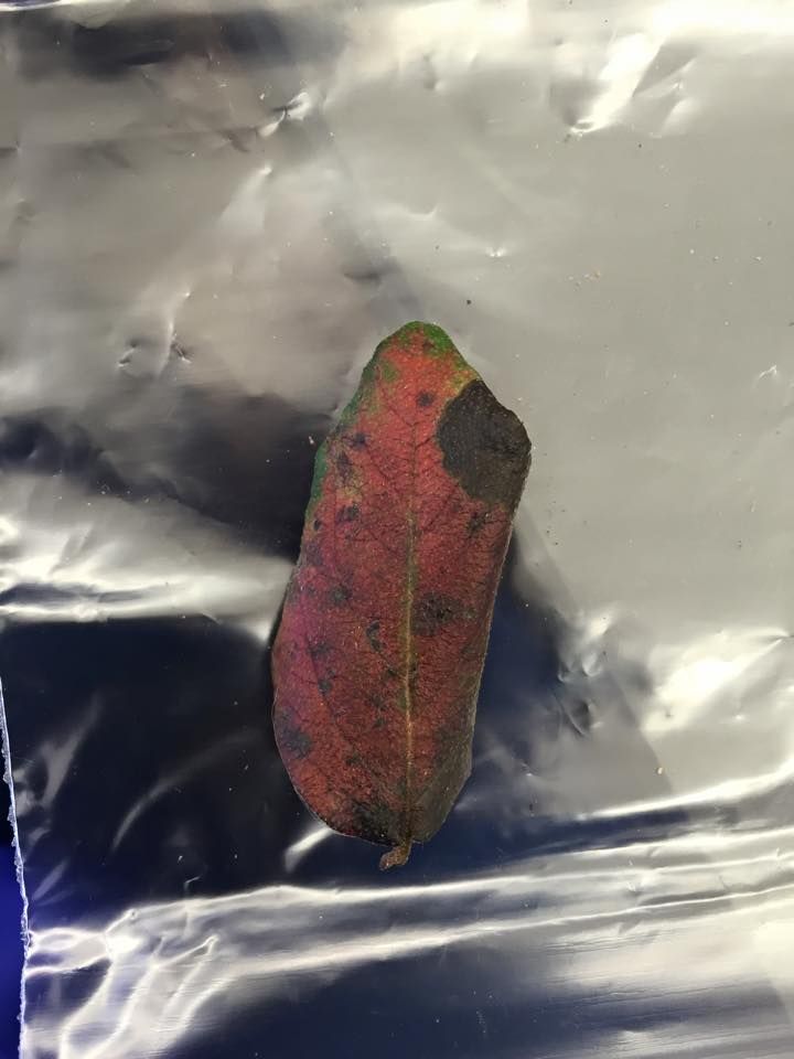
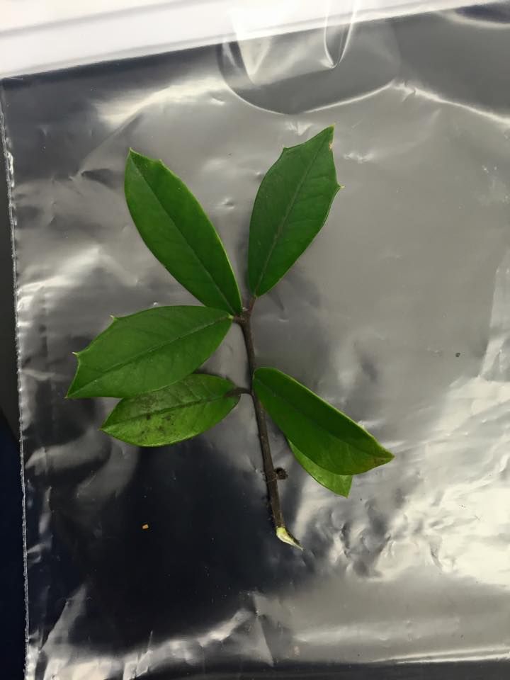
Figure 1: Plant 1 | |||
http://i1382.photobucket.com/albums/ah252/ambujs/10978621_10204583346157117_3996643702295370363_n_zps8531a4a1.jpg | |||
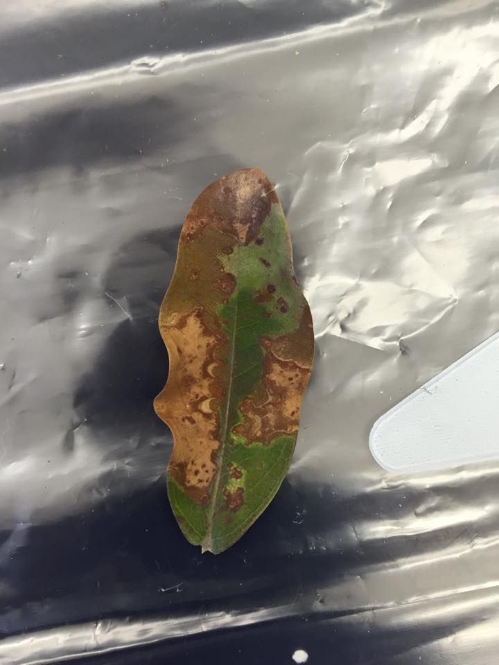
Figure 2: Plant 2 | |||
http://i1382.photobucket.com/albums/ah252/ambujs/10252098_10204583346277120_6932870819414995535_n_zpsb42c5fc9.jpg | |||
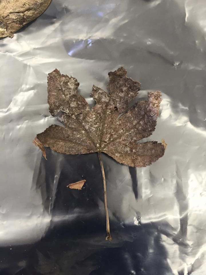
Figure 3: Plant 3 | |||
http://i1382.photobucket.com/albums/ah252/ambujs/10387208_10204583346437124_2035102533959822300_n_zps260ff407.jpg | |||
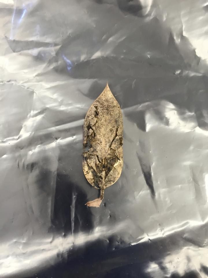
Figure 4: Plant 4 | |||
http://i1382.photobucket.com/albums/ah252/ambujs/10950699_10204583346717131_154076383926522631_n_zps3d13562e.jpg | |||
Figure 5: Plant 5 | |||
http://i1382.photobucket.com/albums/ah252/ambujs/10612791_10204583347077140_4723469001341755998_n_zpsb5b4bc49.jpg | |||
Table 2: Fungi Classification | |||
http://i1382.photobucket.com/albums/ah252/ambujs/Table2Lab4_zpsfb5b6c3f.jpg | |||
Figure 6: Mushroom Sample from Fungi Classification | |||
http://i1382.photobucket.com/albums/ah252/ambujs/10978631_10204583484400573_6703159001730070281_n_zpsb138a8ec.jpg | |||
'''Conclusion:''' | |||
Judging from the varying vascularization of the five leaves identified, it can be concluded that the transect has diverse plant life. From the small sample of leaf litter, five very distinct plant types with few similar characteristics were identified. In addition, the diversity of fungi was observed under the microscope. Most interestingly, sporangia – or site of spores used for asexual reproduction of fungi – were observed in the basidiomycota. For further research, although no seeds were found during the sampling of the transect, they could be retrieved by looking more closely beneath the soil if another sampling were to be prepared. | |||
'''2.10.15''' Very good notebook entry. Could include more detailed description of gram stain methods or include a link to procedure and describe 16s PCR set-up. '''SK''' | |||
== Lab 3 (2/29/15) == | |||
'''Purpose:''' | |||
The purpose of this lab is to test the antibiotic resistance of bacteria found in the hay infusion of transect 3. | |||
'''Materials & Methods:''' | |||
Four diluted samples from the hay infusion were plated onto 8 petri dishes (four with agar and another four with agar plus tetracycline). Over the next week, bacteria on the plate either colonized or died. | |||
Samples of four bacteria were counted (two from the second least dilute tetracycline-positive plate and two from the second least dilute tetracycline-negative) and observed on a wet mount. Additionally, a gram stain was conducted on the four bacteria colonies to determine whether they were gram positive or gram negative. | |||
'''Data & Observations:''' | |||
The hay infusion was settled and more homogenous after two weeks from being created. The niches observed a week prior were now indistinguishable from one another. The infusion also smelled less moldy and about 2 inches of water evaporated. These changes may have been a result of the increasing presence of microorganisms, which expanded their niches to make room for more of themselves. | |||
There were only bacteria present on the eight agar plates after observation. It is likely that archaea are not present in the plates because they only exist in extreme environments. | |||
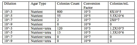
Table 1: | |||
http://i1382.photobucket.com/albums/ah252/ambujs/Table1OWW3_zps5d1be3b6.jpg | |||
Legend: There are many more colonies on plates without tetracycline than those with tetracycline. This difference implies that tetracycline effectively impedes the growth of bacteria. | |||
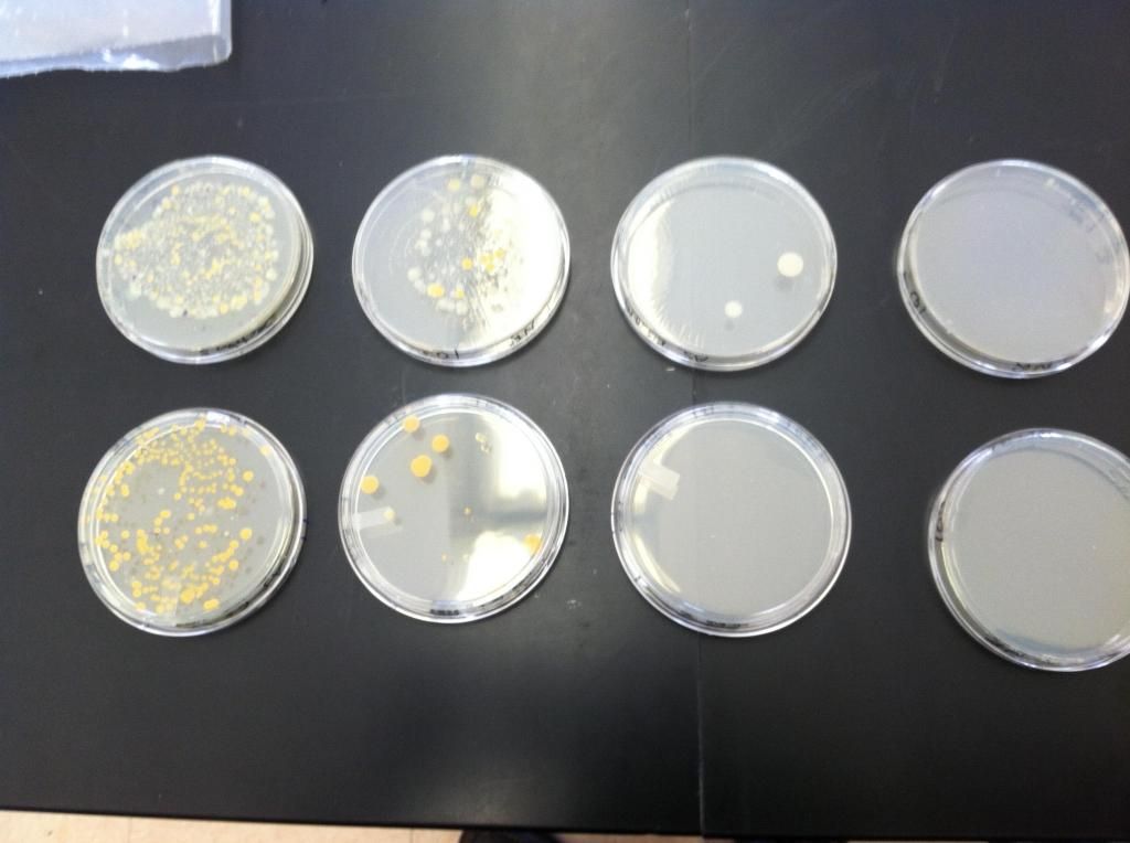
Figure 1: | |||
http://i1382.photobucket.com/albums/ah252/ambujs/Figure1OWW3_zpsd335c991.jpg | |||
Legend: The four plates with tetracycline (above) visibly have more bacteria growing than the four plates without tetracycline (below). In addition, the plates that are less diluted (left) have more bacteria growing than the plates that are more diluted (right). In other words, as dilution of the sample increased, the number of bacteria present on the plates decreased. | |||
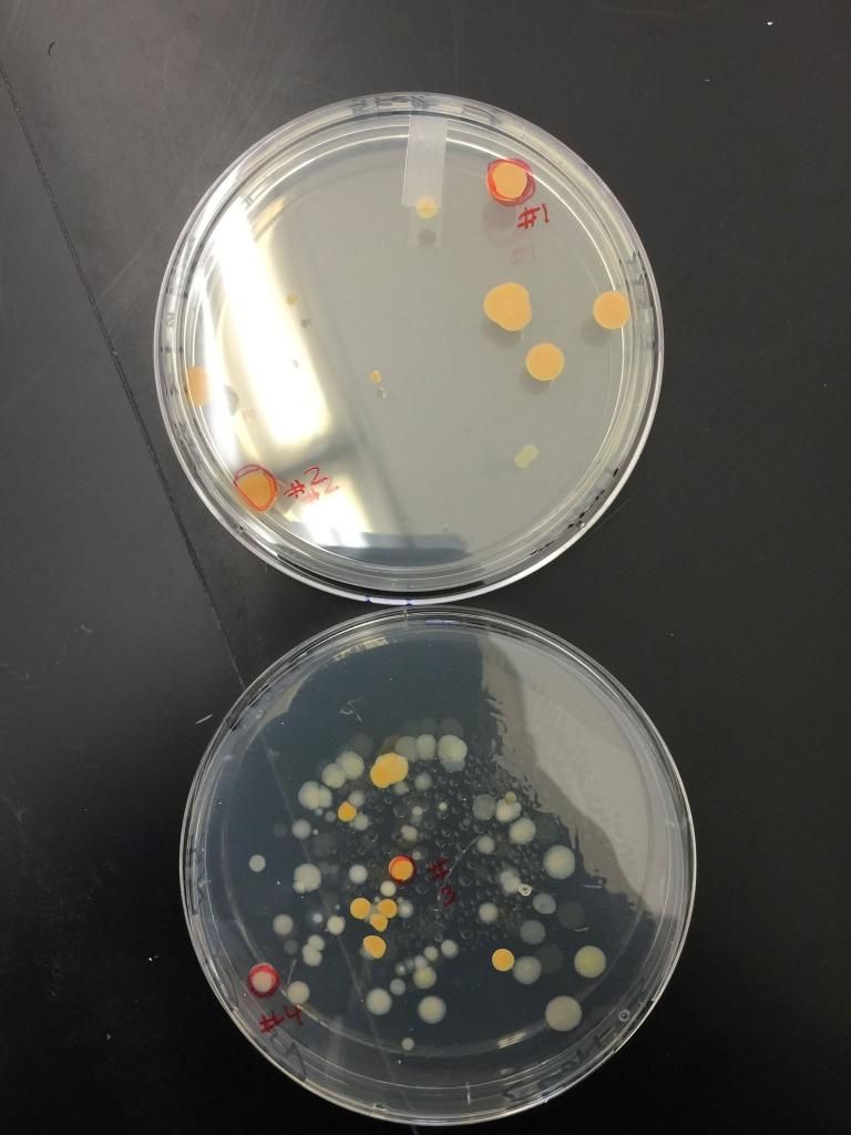
Figure 2: | |||
http://i1382.photobucket.com/albums/ah252/ambujs/Figure2OWW4_zps616a74a4.jpg | |||
Legend: This figure indicates the four colonies of bacteria from the least dilute plates that underwent closer examination in a wet mount and gram stain. They are numbered from 1-4. | |||
Table 2: | |||
http://i1382.photobucket.com/albums/ah252/ambujs/Table2OWW3_zpsdfb0b22e.jpg | |||
Legend: Table 2 indicates the visual descriptions of the four colonies of bacteria from the naked eye and 100x microscopic magnification. Of the colonies observed, three were orange or contained bacillus bacteria. In addition, half of the bacteria observed were motile rather than non-motile. The table also indicates the results of the gram stain. | |||
Figure 3: Colony 1 | |||
http://i1382.photobucket.com/albums/ah252/ambujs/Colony1906691_10153034902323186_7477939883973551244_o_zps0b02865a.jpg | |||
Legend: Figure of Colony 1 in 100x magnification after gram stain. The bacteria appear circular and motile. | |||
Figure 4: Colony 2 | |||
http://i1382.photobucket.com/albums/ah252/ambujs/Colony21514521_10204579697145894_7121853807836957507_n_zpse8150263.jpg | |||
Legend: Figure of Colony 2 in 100x magnification after gram stain. The bacteria appear bacillus and non-motile. | |||
Figure 5: Colony 3 | |||
http://i1382.photobucket.com/albums/ah252/ambujs/Colony310459026_10153034902183186_2164467798252695194_o_zps2c7e33b5.jpg | |||
Legend: Figure of Colony 3 in 100x magnification after gram stain. The bacteria appear bacillus and non-motile (but floating in the water added to the slide). | |||
Figure 6: Colony 4 | |||
http://i1382.photobucket.com/albums/ah252/ambujs/COLONY410923782_10153034902148186_8941320641010632484_o_zps1e9ff908.jpg | |||
Legend: Figure of Colony 1 in 100x magnification after gram stain. The bacteria appear bacillus and motile. | |||
'''Conclusion:''' | |||
Of the eight dishes, those without the tetracycline, the bacteria are widespread and appear orange and white. Without tetracycline, the bacteria are primarily orange with only some white and confined to their own circles. This indicates that the orange species is mostly unaffected by the tetracycline. | |||
Further examination of the bacteria sampled from the tetracycline-positive plates revealed that they were gram positive. By contrast, the bacteria from the tetracycline-negative were gram negative. Therefore, the gram-positive bacteria in the hay infusion are tetracycline resistant since they grew despite in the presence of the antibiotic and gram-negative bacteria are sensitive to tetracycline since they were absent in its presence. | |||
A study by Ian Chopra and Marilyn Roberts conduced in 2001 confirms the gram-negative bacterial sensitivity to tetracycline. The mechanism of action proposed by this and many other studies (Roberts MC, 1996) is active efflux protein that transports the protein out of the bacteria if it were to enter. | |||
'''2.4.15''' Good notebook entry. Well organized but use headings for Purpose, Methods, Observations and Conclusions. '''SK''' | |||
== Lab 2 (1/22/15) == | |||
'''Purpose''' | |||
The purpose of this lab is to identify the different organisms - specifically protists and algae - present in the hay infusion developed from the transect sample from last week. | |||
'''Materials & Methods''' | |||
The hay infusion developed by mixing distilled water, powdered milk, and land samples taken directly from the transect. Organisms were extracted from three different niches in the infusion (bottom, middle, top) using a transfer pipette. They were plated on microscope slides and examined using the 40x objective. The organisms were identified using a dichotomous key. | |||
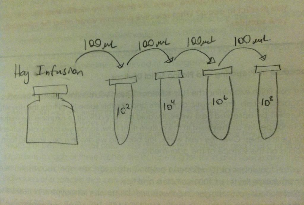
Next, A serial dilution was prepared and plated onto agar plates with and without tetracycline to assess antibacterial resistance of the bacteria in the hay infusion in a week's time. In order to create the serial dilution, 100 microliter sample from the hay infusion was diluted with sterile broth. The figure below illustrates the dilution process. | |||
http://i1382.photobucket.com/albums/ah252/ambujs/7d70b5d4-0854-4498-a1b4-516a4a67ad90_zps72e769c4.jpg | |||
'''Data & Observations''' | |||
The two organisms identified from the top of the hay infusion include chlamydomonas and euplotes sp. Busaria Truncatella and paramecium aurelia were found in the middle. Finally, paramecium bursaria and vorticella were found at the bottom of the hay infusion. | |||
'''Conclusion''' | |||
Upon further examination of the protists extracted, it can be concluded that they are diverse in nature. The organisms differ close versus away from plant matter because those that are far away from plants most likely do not rely on them for food. All of the species are motile (and were therefore hard to locate under the microscope). Paramecium aurelia, paramecium bursaria, bursaria truncatella and euplotes sp are ciliates that rely on cilia for motility whereas vorticella and chlamydomonas have flagellas. Of the six identified, chlamydomonas is the only photosynthetic organism whereas the remaining five rely on external sources of food. Finally, chlamydomonas is the only algae whereas the remaining five are protozoa. | |||
Chlamydomonas are photosynthetic, unicellular organisms that replicate via both sexual and asexual reproduction. Therefore, this organism fits the five necessities of life (energy, cells, replication, information, and evolution). Firstly, they acquire and use energy in the form of sunlight, which is converted to energy through photosynthesis. They are unicellular, and therefore contain at least one membrane bound cell (which comprises the whole organism). They replicate in the form of cell division, and sometimes transfer information creating new daughter cells. As seen in the volvocine line from which chlamydomonas originate, these organisms have also evolved into more complex organisms, such as the volvox. | |||
If the hay infusion “grew” for another two months, then the carrying capacity would steadily decrease. The organisms would reproduce and simultaneously compete for food and space. The competition for food would result in fierce competition as populations grew and food sources dwindled. Therefore, the number of organisms that would be able to survive in each niche would decrease, and only the fit organisms would remain. In this way, the selective pressure of food availability would lead to population change from generation to generation. | |||
== Lab 1 (1/15/15) == | |||
'''Purpose:''' | |||
The purpose of this lab is to examine biodiversity of an assigned transect at American University. | |||
'''Materials & Methods:''' | |||
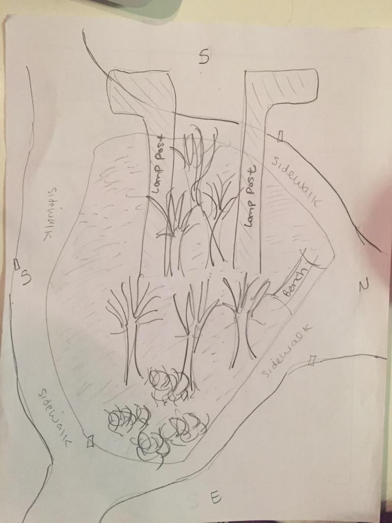
Firstly, the transect assigned transect was located in the northwest corner of American University between Butler Pavilion and Hughes hall. A map was drawn of the 20 by 20 area labeled by Popsicle sticks. The transect was examined for abiotic and biotic features. After recording abiotic and biotic features, a 50 mL sample of soil and leaves was taken from the transect to create a hay infusion to be used in future labs. | |||
'''Data & Observations''' | |||
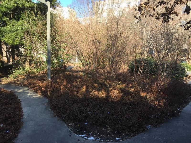
It is approximately 20 by 20 feet containing sidewalk on both sides of a patch of grassy land, containing trees, plants, and lamp posts. | |||
http://i35.photobucket.com/albums/d197/corey92/IMG_8206_zps21726b40.jpg | |||
The five abiotic features observed in the transect include a two metal lamp posts, littered ribbon, sidewalk, pebbles, and a cigarette bud. The five biotic features observed include a small black squirrel, a human between the ages of 18 and 22, seven large trees, several small ferns, and weeds. | |||
http://i1382.photobucket.com/albums/ah252/ambujs/photo_zpsd7c873fd.jpg | |||
'''Conclusion''' | |||
From the various biotic and abiotic features in the transect, it can be concluded that the transect is home to or used by various species that live amongst abiotic features. | |||
== Test post. == | |||
Latest revision as of 21:24, 25 March 2015
Lab 6-8 (2/19/15-3/5/15)
Purpose:
The purpose of this experiment was to determine the effects of caffeine on embryonic development, using zebrafish as a model organism.
Materials & Methods:
Preliminary research was conducted to learn about the possible effects of caffeine on embryonic development of zebrafish. Published papers were used to develop predictions and experimental plan.
Two petri dishes were acquired and labeled as caffeine negative (control) and caffeine positive (experimental). The control environment was prepared by placing 25 mLs of distilled water in a petri dish using a graduated pipette. Additionally, the experimental environment was prepared by placing 25 mLs of 25 mg/L caffeine in the petri dish labeled caffeine positive. 20 healthy zebrafish embryos 24 hours post-fertilization (hpf) were placed in each of the dishes. The embryos were observed under a dissecting microscope to determine developmental stage based on motility, color, and size.
The next day after experimental set-up, dead embryos were removed from each of the petri dishes. In addition, the developmental stage of the living embryos was assessed based on size and physiological features of the embryo.
On the fourth day, 10 mL of solution, debris, and dead fish were removed from each petri dish. To account for removed or evaporated solution, 25 mL of the water and caffeine solutions were added to the control and experimental groups, respectively. The developmental stage of the embryos was recorded.
On the seventh day, the zebrafish were again examined to determine their developmental stage and physiological features. Thereafter 10 mL of caffeine and water were added to account for evaporated water. Additionally, one fish from each of the petri dishes was removed and placed in paraformaldehyde for further examination.
On the eleventh and fourteenth days, debris and dead fish were removed from each petri dish. The approximate developmental stage was recorded along with physical health, such as heart rate, pigmentation, and morphological features. On the fourteenth day, the experiment was terminated by removing dead fish, placing live fish in fresh environments, and discarding the petri dishes. Final observations were recorded and analyzed.
Data & Observations:
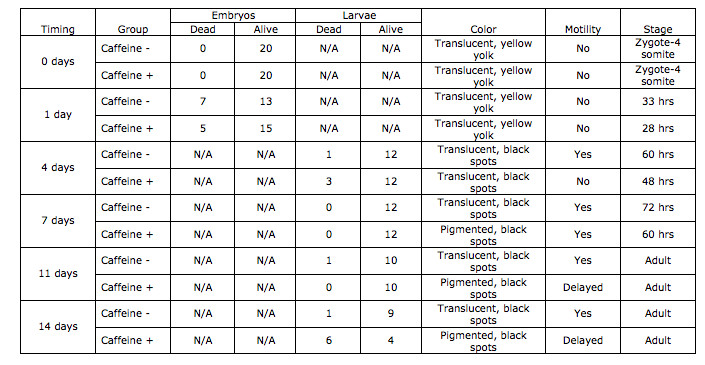
Table 1: Zebrafish Development Over Two Weeks
Legend: Table 1 illustrates the number of zebrafish embryos and hatchings that were dead and alive per observation. It also shows the color, motility and average stage of development of the zebrafish in each group. The zebrafish in the caffeine positive environment were consistently underdeveloped than those in the caffeine negative environment. Additionally, the survival rate of the zebrafish in the caffeine environment is lower than that of the zebrafish in the control environment.
Conclusion
The zebrafish in the experimental group had decreased motility, greater pigmentation, shorter body length, shorter body length, and slowed developmental timelines. In addtion, the number of living zebrafish in the caffeine rich environment was five fish lower than those in the control. Based on these results, it is concluded that caffeine negatively affects embryogenesis in zebrafish relative to the control.
Lab 7 (2/26/15)
Purpose:
The purpose of this experiment is to use DNA sequences to identify species of bacteria found in the transect using polymerase chain reaction (PCR), gel electrophoresis and DNA sequencing to distinguish the 16S gene, which is highly variable.
Materials & Methods:
Samples from a hay infusion were diluted and plated onto nutrient agar and tetracycline treated plates. Bacteria were left to grow on the plates over the next week. Two colonies from each environment were isolated and transferred to 100 microliters of water in four 200 microliter tubes.
The four tubes were incubated at 100 degrees celcius in a heated water bath. After ten minutes, the samples were centrifuged ay 13,400 rpm for 5 minutes. Following centrifugation, 5 microliters of the resulting supernatant fluid was placed with 20 microliters of PCR mixture (including primers and polymerase) in a new labeled tube.
After one week, the PCR product was stained and placed in an agarose gel in a gel electrophoresis apparatus. Using a P-100, the samples were pipetted into wells on the gel along with a ladder. The gel was placed with the well facing the negative charge so that the DNA product would travel to the positive end after initiating the electrical field.
Two of the remaining samples of the PCR product from the tetracycline-rich plates were sent for sequencing. The DNA sequences were then copied and pasted into a Blast software to determine the genetic identity of the bacterial samples.
Data & Observations:
Figure 1:
http://i1382.photobucket.com/albums/ah252/ambujs/IMG_3845_zpsw8floqfb.jpg
Legend: The first well (to the left) contains a ladder that is used to identify the length of the DNA strands whereas the next two wells contain samples of the PCR product with shorter DNA strands.
DNA sequences:
1) MB49 - NNNNNNNNNNNNNNNNANNNTGCAGCCGAGCGGTATTTGTCCTTCGGGACAGAGAGAGCGGCGTACGGGTGCGGAACACG TGTGCAACCTACCTTTATCAGGGGGATAGCCTTTCGAAAGGAAGATTAATACCCCATAATATAAGTCAAGGCATCTTGAT TTATTGAAAACTCCGGTGGATAGAGATGGGCACGCGCAAGATTAGATAGTTGGTAGGGTAACGGCCTACCAAGTCAATGA TCTTTAGGGGGCCTGAGAGGGTGATCCCCCACACTGGTACTGAGACACGGACCAGACTCCTACGGGAGGCAGCAGTGAGG AATATTGGACAATGGGTGAGAGCCTGATCCAGCCATCCCGCGTGAAGGACGACGGCCCTATGGGTTGTAAACTTCTTTTG TATAGGGATAAACCTACTCTCGTGAGAGTANCTGAAGGTACTATACGAATAAGCACCGGCTAACTCCGTGCCAGCAGCCG CGGTAATACGGAGGGTGCAAGCGTTATCCGGATTTATTGGGTTTAAAGGGTCCGTAGGCGGGCTTGTAAGTCAGTGGTGA AATCTCATAGCTTAACTATGAAACTGCCATTGATACTGCAGGTCTTGAGTAAAGTAGAAGTGGCTGGAATAAGTAGTGTA GCGGTGAAATGCATAGATATTACTTANAACACCAATTGCGAAGGCAGGTCACTATGTTTTAACTGACGCTGATGGACGAA AGCGTGGGGAGCGAACAGGATTANATACCCTGGTAGTCCACGCCGTAAACGATGCTNACTCGTTTTTGGGCTTTCGGGTT CAGAGACTAAGCGAAAGTGATAAGTTAGCCACCTGGGGAGTACGTTCGCAAGAATGAAACTCAAAGGAATTGACGGGGGC CCGCACAANCGGTNNTTATGTGGNTTAATTCGATGATANNCGAGGAACCTTANCAAAGGCTNAAATGGGAATTGACAGGN TTANAAAATAGACTTTTCTTCNNACNATTTTCAAGNTGCTGCATGGNNGTCNNCAGCTCGTGCCNTGAGTGTNGNTAAGT CCTGCAACNANCNCAACCCNGNNNNTANNTNNCATNNTTCAGTTNGGGANNNNTAGNNNN
BLAST result: Chryseobacterium sp. LDVH 3 16S ribosomal RNA gene, partial sequence
2) MB50 - NNNNNNNNNNNNNNNNNNNNNANNNNTGCAGCCGAGCGGTATTGTTTCTTCGGAAATGAGAGAGCGGCGTACGGGTGCGG ANCNNNTGTGCAACCTGCCTTTATCTGGGGGATAGCCTTTCGAAAGGGAGATTAATACCCCATAATATATTAAGTGGCAT CACTTGATATTGAAAACTCCGGTGGATAGAGATGGGCACGCGCAAGATTAGATAGTTGGTGAGGTAACGGCTCACCAAGT CTACGATCTTTAGGGGGCCTGAGAGGGTGATCCCCCACACTGGTACTGAGACACGGACCAGACTCCTACGGGAGGCAGCA GTGAGGAATATTGGACAATGGGTGAGAGCCTGATCCAGCCATCCCGCGTGAAGGACGACGGCCCTATGGGTTGTAAACTT CTTTTGTATAGGGATAAACCTACTCTCGTGAGAGTAGCTGAAGGTACTATACGAATAAGCACCGGCTAACTCCGTGCCAG CAGCCGCGGTAATACGGAGGGTGCAAGCGTTATCCGGATTTATTGGGTTTAAAAGGGTCCGTANGCGGATCTGTAAGTCA GTGGTGAAATCTCACAGCTTAACTGTGAAAACTGCCATTGATACTGCAGGTCTTGAGTGTTGTTGAAGTANCTGGAATAA GTAGTGTANCGGTGAAATGGCNTAGATATTACTTAGAAACACCAATTGCNAAGGCTNGTTACTAANCAACAACTGACNCT GATGGACGAAANCGTGGNGGAGCGAACAGGATTANATACCCCTGGNAN
BLAST result: Chryseobacterium sp. StRB028 gene for 16S rRNA, partial sequence
Conclusions:
After analyzing the gel, the presence of 16S gene can be concluded because the DNA that was processed using PCR had shorter strands, supposedly cut at the site of the 16S gene.
The 16S gene is highly variable and certain sequences are specific to different types of bacteria. For this purpose, after confirming the presence of the 16S gene in the bacteria, the identity of the bacteria in the transect can determined from their sequence.
The 16S gene showed that the identity of the tetracycline-resistant sample is Chryseobacterium. Chryseobacterium are generally yellow-raised colonies with rod-shaped bacteria, which matches the morphology of the bacteria found on the plates. In addition, they are generally gram negative. This is contrary to the results of the gram stain of the tetracycline-resistant bacteria, which were gram positive. This discrepancy is possibly due to false interpretation of the gram stains in previous experiments. In hindsight, this is consistent with the fact that the gram stain resulted in stains of bacteria that were intermediate between pink and purple. Therefore, the results stains were difficult to ascertain. Finally, the Chryseobacterium genus contains members that are both motile and non-motile. This is consistent with the results of the observation of the bacteria colonies in 100x perspective, which showed both motile and non-motile colonies.
2.20.15
Very good lab book entry. Some good description in conclusions. (transect was 20 feet x 20 feet, not meters).
Decent food web but could be more detailed.
SK
Lab 5 (2/12/15)
Purpose:
The purpose of this lab is to identify the invertebrates living in the transect. In addition, the interactions between organisms – invertebrates and vertebrates – living in the transect are theorized.
Materials & Methods:
Leaf litter and ground soil were collected from the transect. Back in the lab, a 25 mL solution of half water and half ethanol was prepared and placed in a plastic tube. Screening material was taped to the inside of a glass funnel, which was filled with the transect sample. After placing the funnel into the ethanol solution, it was placed under a 40 watt lamp on a lab bench.
Over the course of a week, any invertebrates that were in the sample fell to the bottom of the ethanol solution to be preserved. The ethanol solution containing invertebrates was then poured into two petri dishes to be observed under a microscope. The samples were checked for invertebrates to be identified.
Groups of vertebrates that inhabit the transect were observed and classified. After determining classification of each vertebrate species, the biotic and abiotic features from which they would benefit were listed. Finally, a food web was modeled for the various organisms in the transect.
Data & Observations:
http://i1382.photobucket.com/albums/ah252/ambujs/Table1Invertebrates_zpsb6755f03.jpg
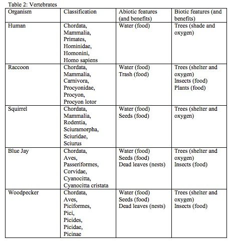
http://i1382.photobucket.com/albums/ah252/ambujs/Table2Vertebrates_zps7ea15382.jpg
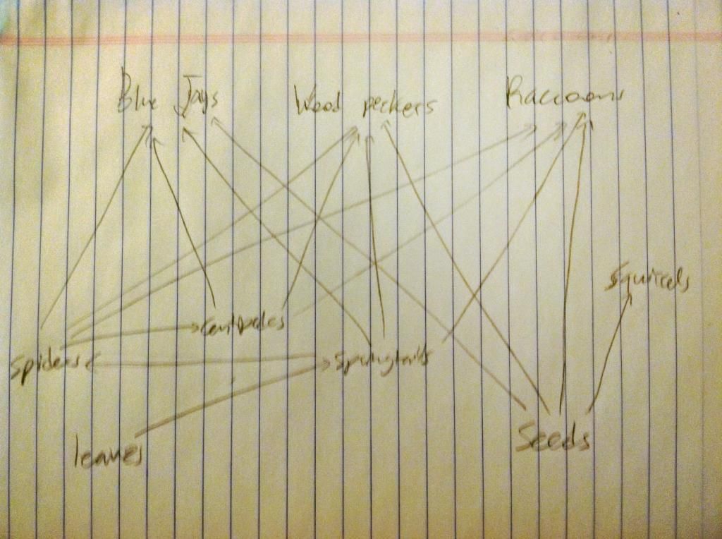
Figure 1:
Conclusion:
There was a variety of small invertebrates found in the transect. The invertebrates extracted from the Berlese Funnel ranged from 1 to 7 mm. The smallest organism was the ground spider whereas the largest was the centipede. Additionally, springtails were most abundant as more than one was found in the selected sample. Since four invertebrates were found from the small sample of soil and leaves, it can be concluded that the transect as a whole hosts a plethora of small invertebrates of different types that feed on each other and their surroundings.
Additionally, five types of chordata were observed in the transect. These vertebrates are ecologically interrelated to the invertebrates as demonstrated by the food web (see figure 1). Together, these organisms represent the concept of community because they interact with each other within the transect. For example, the birds prey on the invertebrates whereas the squirrels and raccoons both compete for the available food resources, such as seeds. This relates to the concept of carrying capacity because there is a limited availability of resources, such as food, shelter, and space, in the 20 by 20 meter zone. In other words, there is a maximum number of individuals that can inhabit the space at a certain period of time. Finally, the trophic levels of these individuals should be considered. Raccoons and birds are large animals that are able to consume smaller species, such as the invertebrates identified above. This places them in a different trophic level than the invertebrates, who primarily feed on leaves and other creatures their size or smaller.
2.20.15 Very good notebook entry. Detailed descriptions and well organized. SK
Lab 4 (2/5/15)
Purpose:
The purpose of this lab is to examine the diversity of plants that exist in Transect 3 as well as observed fungi.
Materials & Methods:
Two Ziploc bags were filled with samples from the transect. The first was 20 percent soil and 80 percent groundcover. The second was filled with leaves from the plants in the transect, both dead and alive. Back in the lab, five of the leaves in the second bag were chosen for further examination. They were observed for general appearance and vascularization. Second, fungi were observed under dissecting microscopes and classified as zycomycata, basidiomycata, and ascomycata based on observable characteristics. Finally, the remaining sample of leaves and soil was placed in a funnel above ethanol to extract invertebrates over the course of a week. This berlese funnel will be examined in the next lab.
Data & Observations:
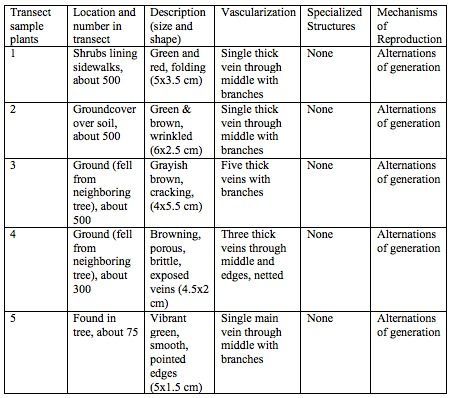
Table 1: Plant Characterization
http://i1382.photobucket.com/albums/ah252/ambujs/Table1Lab4_zpsdde6268a.jpg
Figure 1: Plant 1
Figure 2: Plant 2
Figure 3: Plant 3
Figure 4: Plant 4
Figure 5: Plant 5
Table 2: Fungi Classification
http://i1382.photobucket.com/albums/ah252/ambujs/Table2Lab4_zpsfb5b6c3f.jpg
Figure 6: Mushroom Sample from Fungi Classification
Conclusion:
Judging from the varying vascularization of the five leaves identified, it can be concluded that the transect has diverse plant life. From the small sample of leaf litter, five very distinct plant types with few similar characteristics were identified. In addition, the diversity of fungi was observed under the microscope. Most interestingly, sporangia – or site of spores used for asexual reproduction of fungi – were observed in the basidiomycota. For further research, although no seeds were found during the sampling of the transect, they could be retrieved by looking more closely beneath the soil if another sampling were to be prepared.
2.10.15 Very good notebook entry. Could include more detailed description of gram stain methods or include a link to procedure and describe 16s PCR set-up. SK
Lab 3 (2/29/15)
Purpose:
The purpose of this lab is to test the antibiotic resistance of bacteria found in the hay infusion of transect 3.
Materials & Methods:
Four diluted samples from the hay infusion were plated onto 8 petri dishes (four with agar and another four with agar plus tetracycline). Over the next week, bacteria on the plate either colonized or died.
Samples of four bacteria were counted (two from the second least dilute tetracycline-positive plate and two from the second least dilute tetracycline-negative) and observed on a wet mount. Additionally, a gram stain was conducted on the four bacteria colonies to determine whether they were gram positive or gram negative.
Data & Observations:
The hay infusion was settled and more homogenous after two weeks from being created. The niches observed a week prior were now indistinguishable from one another. The infusion also smelled less moldy and about 2 inches of water evaporated. These changes may have been a result of the increasing presence of microorganisms, which expanded their niches to make room for more of themselves.
There were only bacteria present on the eight agar plates after observation. It is likely that archaea are not present in the plates because they only exist in extreme environments.
Table 1:
http://i1382.photobucket.com/albums/ah252/ambujs/Table1OWW3_zps5d1be3b6.jpg
Legend: There are many more colonies on plates without tetracycline than those with tetracycline. This difference implies that tetracycline effectively impedes the growth of bacteria.
Figure 1:
http://i1382.photobucket.com/albums/ah252/ambujs/Figure1OWW3_zpsd335c991.jpg
Legend: The four plates with tetracycline (above) visibly have more bacteria growing than the four plates without tetracycline (below). In addition, the plates that are less diluted (left) have more bacteria growing than the plates that are more diluted (right). In other words, as dilution of the sample increased, the number of bacteria present on the plates decreased.
Figure 2:
http://i1382.photobucket.com/albums/ah252/ambujs/Figure2OWW4_zps616a74a4.jpg
Legend: This figure indicates the four colonies of bacteria from the least dilute plates that underwent closer examination in a wet mount and gram stain. They are numbered from 1-4.
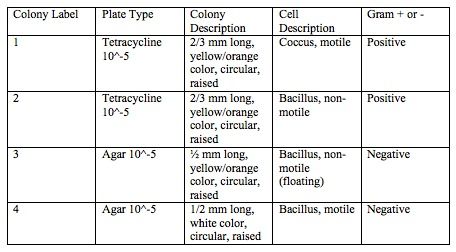
Table 2:
http://i1382.photobucket.com/albums/ah252/ambujs/Table2OWW3_zpsdfb0b22e.jpg
Legend: Table 2 indicates the visual descriptions of the four colonies of bacteria from the naked eye and 100x microscopic magnification. Of the colonies observed, three were orange or contained bacillus bacteria. In addition, half of the bacteria observed were motile rather than non-motile. The table also indicates the results of the gram stain.
Figure 3: Colony 1
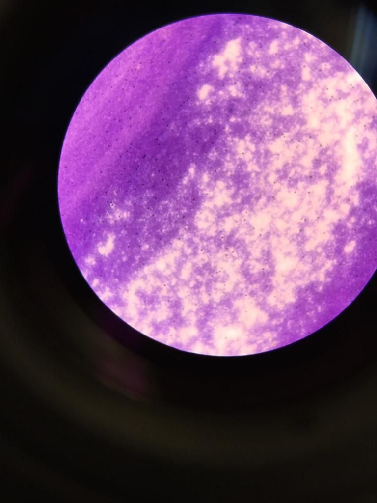
Legend: Figure of Colony 1 in 100x magnification after gram stain. The bacteria appear circular and motile.
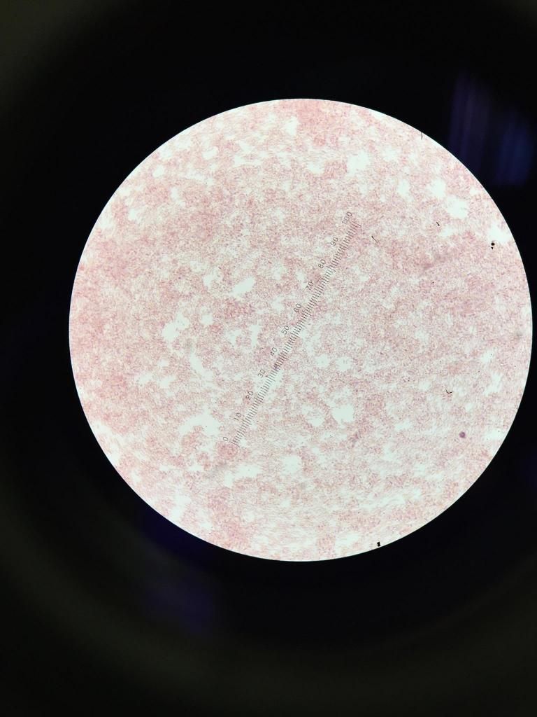
Figure 4: Colony 2
Legend: Figure of Colony 2 in 100x magnification after gram stain. The bacteria appear bacillus and non-motile.
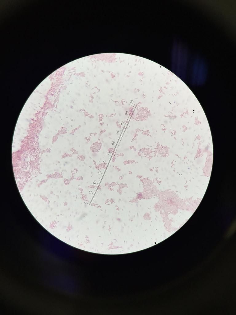
Figure 5: Colony 3
Legend: Figure of Colony 3 in 100x magnification after gram stain. The bacteria appear bacillus and non-motile (but floating in the water added to the slide).
Figure 6: Colony 4
Legend: Figure of Colony 1 in 100x magnification after gram stain. The bacteria appear bacillus and motile.
Conclusion:
Of the eight dishes, those without the tetracycline, the bacteria are widespread and appear orange and white. Without tetracycline, the bacteria are primarily orange with only some white and confined to their own circles. This indicates that the orange species is mostly unaffected by the tetracycline.
Further examination of the bacteria sampled from the tetracycline-positive plates revealed that they were gram positive. By contrast, the bacteria from the tetracycline-negative were gram negative. Therefore, the gram-positive bacteria in the hay infusion are tetracycline resistant since they grew despite in the presence of the antibiotic and gram-negative bacteria are sensitive to tetracycline since they were absent in its presence.
A study by Ian Chopra and Marilyn Roberts conduced in 2001 confirms the gram-negative bacterial sensitivity to tetracycline. The mechanism of action proposed by this and many other studies (Roberts MC, 1996) is active efflux protein that transports the protein out of the bacteria if it were to enter.
2.4.15 Good notebook entry. Well organized but use headings for Purpose, Methods, Observations and Conclusions. SK
Lab 2 (1/22/15)
Purpose
The purpose of this lab is to identify the different organisms - specifically protists and algae - present in the hay infusion developed from the transect sample from last week.
Materials & Methods
The hay infusion developed by mixing distilled water, powdered milk, and land samples taken directly from the transect. Organisms were extracted from three different niches in the infusion (bottom, middle, top) using a transfer pipette. They were plated on microscope slides and examined using the 40x objective. The organisms were identified using a dichotomous key.
Next, A serial dilution was prepared and plated onto agar plates with and without tetracycline to assess antibacterial resistance of the bacteria in the hay infusion in a week's time. In order to create the serial dilution, 100 microliter sample from the hay infusion was diluted with sterile broth. The figure below illustrates the dilution process.
Data & Observations
The two organisms identified from the top of the hay infusion include chlamydomonas and euplotes sp. Busaria Truncatella and paramecium aurelia were found in the middle. Finally, paramecium bursaria and vorticella were found at the bottom of the hay infusion.
Conclusion
Upon further examination of the protists extracted, it can be concluded that they are diverse in nature. The organisms differ close versus away from plant matter because those that are far away from plants most likely do not rely on them for food. All of the species are motile (and were therefore hard to locate under the microscope). Paramecium aurelia, paramecium bursaria, bursaria truncatella and euplotes sp are ciliates that rely on cilia for motility whereas vorticella and chlamydomonas have flagellas. Of the six identified, chlamydomonas is the only photosynthetic organism whereas the remaining five rely on external sources of food. Finally, chlamydomonas is the only algae whereas the remaining five are protozoa.
Chlamydomonas are photosynthetic, unicellular organisms that replicate via both sexual and asexual reproduction. Therefore, this organism fits the five necessities of life (energy, cells, replication, information, and evolution). Firstly, they acquire and use energy in the form of sunlight, which is converted to energy through photosynthesis. They are unicellular, and therefore contain at least one membrane bound cell (which comprises the whole organism). They replicate in the form of cell division, and sometimes transfer information creating new daughter cells. As seen in the volvocine line from which chlamydomonas originate, these organisms have also evolved into more complex organisms, such as the volvox.
If the hay infusion “grew” for another two months, then the carrying capacity would steadily decrease. The organisms would reproduce and simultaneously compete for food and space. The competition for food would result in fierce competition as populations grew and food sources dwindled. Therefore, the number of organisms that would be able to survive in each niche would decrease, and only the fit organisms would remain. In this way, the selective pressure of food availability would lead to population change from generation to generation.
Lab 1 (1/15/15)
Purpose:
The purpose of this lab is to examine biodiversity of an assigned transect at American University.
Materials & Methods:
Firstly, the transect assigned transect was located in the northwest corner of American University between Butler Pavilion and Hughes hall. A map was drawn of the 20 by 20 area labeled by Popsicle sticks. The transect was examined for abiotic and biotic features. After recording abiotic and biotic features, a 50 mL sample of soil and leaves was taken from the transect to create a hay infusion to be used in future labs.
Data & Observations
It is approximately 20 by 20 feet containing sidewalk on both sides of a patch of grassy land, containing trees, plants, and lamp posts.
http://i35.photobucket.com/albums/d197/corey92/IMG_8206_zps21726b40.jpg
The five abiotic features observed in the transect include a two metal lamp posts, littered ribbon, sidewalk, pebbles, and a cigarette bud. The five biotic features observed include a small black squirrel, a human between the ages of 18 and 22, seven large trees, several small ferns, and weeds.
http://i1382.photobucket.com/albums/ah252/ambujs/photo_zpsd7c873fd.jpg
Conclusion
From the various biotic and abiotic features in the transect, it can be concluded that the transect is home to or used by various species that live amongst abiotic features.