User:Alison M. Taylor/Notebook/Biology 210 at AU: Difference between revisions
No edit summary |
No edit summary |
||
| Line 1: | Line 1: | ||
'''Analyzing Invertebrates from the Berlese Funnel''' | |||
'''February 10, 2016''' | |||
[[Image:BerleseFunnelAMT.jpg]] | |||
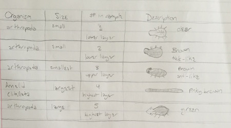
Table 1: Shows data on the five invertebrates collected from transect 2. | |||
The size ranged from microscopic arthropods to an earthworm about two inches long. The organism that was most frequently observed was in the phylum arthropoda and looked like an ant. The sample from the higher layer of the ethanol appeared to contain only these organisms. They were not observed in the second sample from the lower layer, which had less abundant but more diverse arthropods. | |||
'''Characterizing Plants from Transect 2''' | '''Characterizing Plants from Transect 2''' | ||
Revision as of 10:05, 13 February 2016
Analyzing Invertebrates from the Berlese Funnel
February 10, 2016
Table 1: Shows data on the five invertebrates collected from transect 2.
The size ranged from microscopic arthropods to an earthworm about two inches long. The organism that was most frequently observed was in the phylum arthropoda and looked like an ant. The sample from the higher layer of the ethanol appeared to contain only these organisms. They were not observed in the second sample from the lower layer, which had less abundant but more diverse arthropods.
Characterizing Plants from Transect 2
February 3, 2016
Plant collection and observation:
The condition of transect 2 was flooded as it was very rainy and around 48 F. The ground matter and debris were therefore very wet. The samples collected, shown below, were all angiosperms.
Fig. 1 Shows five plant samples selected from Transect 2.
Table 1 Shows characteristics of plant samples collected from the transect.
Observing Fungi:
Sporangia serve as the location for where spores are formed, which is how fungi reproduce, making sporangia vital.
Fig. 2 This sample in the lab is clearly a fungus due to the distinct characteristic of hyphae. This mushroom belongs to basidiomycota.
AMT
Observing Microorganisms via Morphology and Gram Staining
January 26, 2016
Hay Infusion Culture: Final Observation
The hay infusion culture has evaporated roughly an inch since the previous week. The plant matter has further decayed, leaving most of the water a little clearer. The water surface was perhaps more coagulated. The jar still smells like decay, and somewhat like feces. The smell and appearance may change week to week because the organic matter inside will have decomposed further as time goes on. Also, the growth of more bacteria and mold may add to the smell. There will not be any Archaea species growing in the culture though, as the conditions are not extreme enough.
Quantifying Microorganisms:
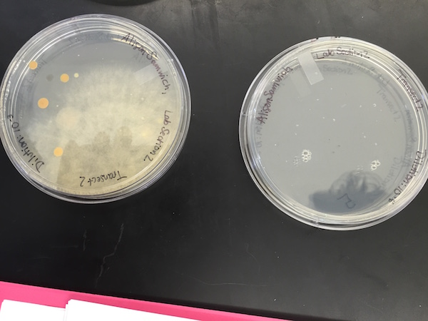
Table 1: Shows the number of colonies of bacteria grown on an agar plate with and without tetracycline, at various dilution levels.
The plates without tetracycline clearly grew many more bacteria than those with the antibiotic did. The most successful growth on a plate with tetracycline was that of mold, so antibiotic is less effective on fungi than bacteria (makes sense). The main kind of bacteria that grew on the non-antibiotic plates is the same type that showed up on the antibiotic plates. A few purple bacteria showed up on the non-antibiotic plates which did not grow at all on the tetracycline plates, indicating that the tetracycline has a greater effect on certain types of bacteria. None of the bacteria seemed to be totally unaffected by tetracycline though, given the lack of growth compared to non-tetracycline plates. The antibiotic Tetracycline hinders bacterial ribsomes from interacting with aminoacyle-tRNA, which prevents protein synthesis in the bacteria NCBI. Tetracycline affects bacteria including E. Coli, Streptococcal pneumoniae, and Bacillus anthracis (anthrax) Tulane PharmWiki
Cell Descriptions:
Table 2: Shows information about each of the four bacterial colonies observed
Figure 1: Drawings of each of the four colonies observed, at 40x magnification.
Figures 1-5: Photographs of all eight agar plates, showing the growth (or lack of) of bacteria and fungi on each.
Citations
Chopra, Ian, and Marilyn Roberts. “Tetracycline Antibiotics: Mode of Action, Applications, Molecular Biology, and Epidemiology of Bacterial Resistance.” Microbiology and Molecular Biology Reviews 65.2 (2001): 232–260. PMC. Web. 29 Jan. 2016.
AMT
Culture Observations and Serial Dilution
January 20, 2016
Figure 1: Serial dilution procedure using hay infusion culture and agar plates
Hay Infusion Culture Observations:
The hay infusion culture's appearance was swampy, clouded water, filled with organic debris. The smell was foul, distinctly like decomposition. There was no apparent life, except for possibly a small amount of mold growing on the jar right above the water line. A sample collected from the bottom revealed a colpidium (70um), paramacium (35um), and paranema (30um). A sample from nearer the top of the water contained volvox (400um), didinium (100um), and vorticella (50um). It is not surprising that this second sample contained protists, as they were drawn from near plant matter, which could serve as nutrition. Still, all six organisms observed were motile, nutrient-consuming protists.
The paramecium observed meets all fundamental characteristics of life. 1) Energy: paramecium acquire energy by consuming nutrients, like the dead plant matter in the hay infusion. 2) Cells: Paramecium are single-celled organisms. They have macronuclei, which are responsible for duties like digestion and protein synthesis. 3) Information: Paramecium's genetic information is replicated during binary fission or conjugation. 4) Replication: Paramecium replicate asexually through binary fission or sexually by conjugation. 5) Evolution: Paramecium's replication method of conjugation is an example of evolving to sexual dimorphism.
If the hay infusion culture was observed after another two months, I hypothesize that there would be more growth, as protists continued to eat decaying plant matter and reproduce, and algae, if any was present, continued to photosynthesize.
Figure 2: Protists observed from samples of hay infusion culture from transect 2.
Examining a Transect at AU & Hay Infusion Culture
January 13, 2016
Purpose: The purpose of this lab was to observe and collect samples from a 20x20ft transect, and then use the samples in a Hay Infusion Culture. This was done to enrich any microorganisms present in the sample. Our hypothesis is that there will be growth in the culture, particularly given that we included samples from still water, where there are likely to be living organisms.
Materials and Methods:
- Observe and draw aerial view of transect 2, just south of Hughes Hall on AU's campus.
- List abiotic and biotic components of the transect.
- Collect various samples, including soil, leaves, and water.
- In the laboratory, add 10.4g of sample ground matter and 6.5 mL of sampled water to a labeled plastic jar.
- Add 500 mL of room-temperature Deerpark water and .1g of dried milk to the jar.
- Cap the jar and shake to mix thoroughly.
- Take the lid off and let it sit for one week.
Data and Observations:
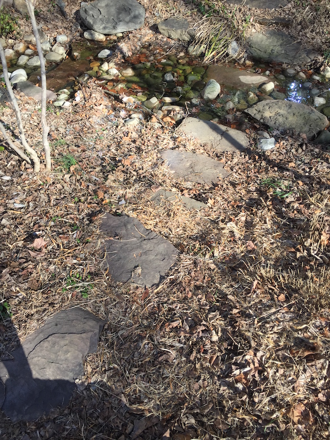
The transect sloped downwards to the west, in the direction the stream was flowing. The water flow was moderately paced, and the outer edges were still. The weather was 28F, sunny, and windy. The biotic components found in the transect were leaves, bushes, trees, plant bulbs, squirrels, and birds. The abiotic elements were rocks, trash (paper, glass bottles), an irrigation control box, water, and soil.
Figure 1: Aerial Drawing of transect 2.
Figure 2: Aerial picture of portion of transect 2.
Figure 3: Panoramic view of entire transect 2.
Conclusions and Future Directions:
So far there are no conclusions to be drawn, but it is known that some of the samples were abiotic and still living, despite the cold temperatures. Next week the Hay Infusion Culture will be observed to see if any protists are present.
AMT


