User:Alison D. Bunnen/Notebook/Biology 210 at AU: Difference between revisions
No edit summary |
No edit summary |
||
| Line 52: | Line 52: | ||
Figure 3. is of Organism 3. In lab we established this organism to be Euglena, however after extensive google image searches, I believe it to be Fusarium graminearum. It is ~50μm, peapod/canoe shaped, long, thin, some are segmented inside, and colorless. | Figure 3. is of Organism 3. In lab we established this organism to be Euglena, however after extensive google image searches, I believe it to be Fusarium graminearum. It is ~50μm, peapod/canoe shaped, long, thin, some are segmented inside, and colorless. | ||
Figure 4. | Figure 4. [[Image:Organism4.jpg]] | ||
Figure 4. is of Organism 4. In lab we could not determine the genus and species. It is ~75μm, disc-like, pods in the middle and colorless. | |||
''IV. Conclusions:'' | ''IV. Conclusions:'' | ||
Revision as of 19:13, 29 January 2015
1/28/15 Lab 3: Microbiology and Identifying Bacteria with DNA Sequences
I. Purpose:
II. Materials and Methods:
III. Data and Observations:
IV. Conclusions:
1/21/15 Lab 2: Identifying Algae and Protists
I. Purpose: The purpose of this lab is to examine and understand the characteristics of algae and protists from my transect.
II. Materials and Methods:
· Make a wet mount from the niche at the top of the jar and make a wet mount from the niche at the bottom of the jar
· View at 4x, 10x, and 40x.
· Make observations of at least 3 different organisms from each niche
· Use a dichotomous key to identify the organisms
III. Data and Observations: My Hay Infusion Culture has a foul smell, almost like rotten garbage. The top of the jar has a layer of film, maybe mold, with a gray/green color. The water is yellow/brown tinged. The bottom of the jar has a thin layer of dirt on it.
Figure 1. is the top view of my Hay Infusion Culture for Group 3
Figure 2. is the side view of my Hay Infusion Culture for Group 3
The organisms below were found in the top of the jar niche.
Figure 1. is of Organism 1. In lab we established this organism to be a Didinium Cyst, however after further research, that is not what Organism 1 is. At the moment, Organism 1 is still unknown. It is ~30μm , almost circular with two indentations at the top and bottom of the cell, little oval specks inside moving, and colorless.
Figure 2. is of Organism 2. In lab we established this organism to be Colpidium. The organism varied in size, one I measured was ~50μm, packman-like face, spins, type of motility undetected, and colorless.
Figure 3. is of Organism 3. In lab we established this organism to be Euglena, however after extensive google image searches, I believe it to be Fusarium graminearum. It is ~50μm, peapod/canoe shaped, long, thin, some are segmented inside, and colorless.
Figure 4. is of Organism 4. In lab we could not determine the genus and species. It is ~75μm, disc-like, pods in the middle and colorless.
IV. Conclusions:
--Alison D. Bunnen 18:33, 29 January 2015 (EST)
1/14/15 Lab 1: Biological Life at AU
I. Purpose: The purpose of today's lab was to observe and record characteristics of an assigned transect. We took samples from our niche to create a Hay Infusion Culture. I hypothesize the transects have copious amounts of bacteria living in the niche. If water is added to the gathered materials and is allowed to incubate, then the bacteria from the niche (in the soil) will grow in the new ecosystem created.
II. Materials and Methods:
· 20 x 20 meter transect to study
· Use a sterile 50 mL conical tube to gather materials such as soil and vegetation from transect
· Place 10-12 grams of gathered materials into a large jar
· Add 500 mLs of deerpark water to the jar
· Add 0.1 gm of dried milk into the jar and screw on lid
· Mix contents gently for 10 seconds
· Place the jar in designated lab section area with the jar labeled and lid off
III. Data and Observations: My assigned transect was group 3: Tall Bushes. I recorded macroscopic observations of abiotic and biotic components in my transect. Then my group made a Hay Infusion Culture from materials gathered from our niche. The Hay Infusion Culture was a mix of ~10 grams of soil and vegetation from my transect, 500 mL of deerpark water, and 0.1 gm of dried milk.
The abiotic components inside the transect included:
· Light
· Water/snow
· Concrete
· A metal lamp post
· Dirt
· Rocks
The biotic components inside the transect included:
· Grass
· Shrub
· Bushes
Figure 1. is an aerial-view diagram of my transect labeled with it's abiotic and biotic components.
Figure 2. is a satellite view of Group 3's transect.
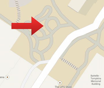
Figure 3. is a map view of Group 3's transect.
IV. Conclusions: There are abiotic and biotic components in Group 3's transect. I will not know if my hypothesis is correct until further examination of the incubated Hay Infusion Culture is done.
--Alison D. Bunnen 22:24, 26 January 2015 (EST)


