CAMRI:HowToScan: Difference between revisions
No edit summary |
No edit summary |
||
| Line 58: | Line 58: | ||
[[Image:qa29-Jul-2015_Page_1.jpg | | [[Image:qa29-Jul-2015_Page_1.jpg | 900px|Siemens EPI product sequence with GRAPPA 3, TR = 2 seconds]] | ||
[[Image:qa29-Jul-2015_Page_2.jpg | | [[Image:qa29-Jul-2015_Page_2.jpg | 900px|MGH Blipped CAIPI multi-band EPI with SMS 2, GRAPPA 2, TR = 1.5 seconds]] | ||
[[Image:qa29-Jul-2015_Page_3.jpg | | [[Image:qa29-Jul-2015_Page_3.jpg | 900px|MGH Blipped CAIPI multi-band EPI with SMS 2, GRAPPA 3, TR = 1.5 seconds]] | ||
Revision as of 12:02, 13 August 2015
|
About CAMRI's Siemens ScannersThe BCM CAMRI has 3 whole-body 3T Siemens Magnetom Trios, labelled 3T-3, 3T-4 and 3T-5. 3T-3 and 3T-4 currently have 8 receive channels, 3T-5 has 32 channels. First, register the patient. A shortcut is to bring up a previously registered patient, and then edit that record. Response RecordingResponses are recorded with a fORP system. Link to Jung Hwan's user guide here: fORP Configuration Visual Stimulus Displays3T-3 and 3T-5 display visual stimuli on direct view LCD screens. Link to manual here: Cambridge Research Systems BOLD Screen User Guide The BOLDScreen has a built-in color profile that can make images appear too dark. Instead, use the default color profile as follows:
Ricky Savjani has redone the gamma calibration on the 3T-5 BOLDscreen, this can be accessed in PsychToolbox as follows. First, Click here to load the table % load in the gamma table
load CRS_32BOLDSCREEN/gammaTable_Spline_fit.mat
% after opening a window, call this to load the load the gamma table to to your Screen
Screen('LoadNormalizedGammaTable', win, gammaTable2*[1 1 1]);
Now, desired intensity levels will be mapped appropriately to the screen. Rough calculation of image size in degrees of visual angle: roughly 76.5 cm from screen to eyeball height of screen is 13.7 in = 34. 8 cm width of screen is 34.8 * (1024/768) = 46.4 cm atan(23.2/76.5) = 0.29 rad = 16.6 degrees ~30 degrees full width Misc NotesSlice OrderingDavid Ress' former student Andrew Florens examined the acquisition time stamps and found that axial slices are collected from Superior to Inferior (head to foot). If an odd number of slices is acquired, the slice order is 1, 3, 5, 7, etc. 2, 4, 6, 8 etc. If an even number of slices is acquired, the slice order is reversed: 2, 4, 6, 8, etc. 1, 3, 5, 7, etc.
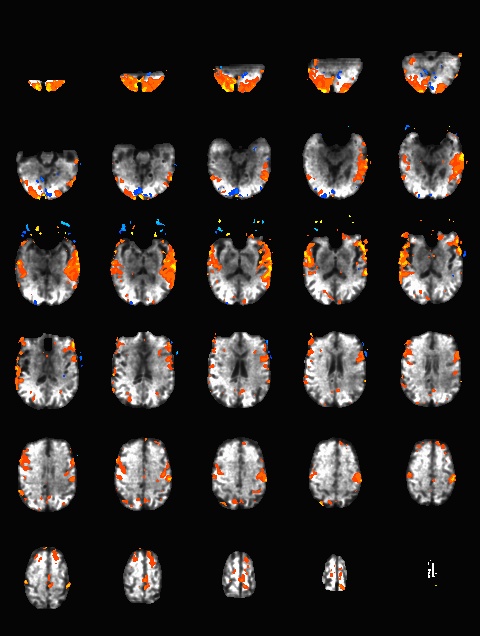
What pulse sequence to use for fMRIThere are a variety of different pulse sequences that can be used for fMRI, including the standard Siemens EPI product sequences; multi-band EPI sequences from MGH and Univ. of Minnesota; and spiral sequences implemented by David Ress.
Similar results were obtained with a phantom: |


