Biomod/2015/Kansai/Sources: Difference between revisions
No edit summary |
No edit summary |
||
| (3 intermediate revisions by the same user not shown) | |||
| Line 9: | Line 9: | ||
<table border="1" cellpadding="4" width="1000" > | <table border="1" cellpadding="4" width="1000" > | ||
<tr align="center"> | <tr align="center"> | ||
<td width="180" bgcolor="#800000">[[Biomod/2015/Kansai|<font face="FANTASY,cursive,Arial" color="white"> | <td width="180" bgcolor="#800000">[[Biomod/2015/Kansai|<font face="FANTASY,cursive,Arial" color="white">HOME</font>]] </td> | ||
<td width="180" bgcolor="#000000">[[Biomod/2015/Kansai/Team|<font face="FANTASY,cursive,Arial" color="white">Team</font>]] </td> | <td width="180" bgcolor="#000000">[[Biomod/2015/Kansai/Team|<font face="FANTASY,cursive,Arial" color="white">Team</font>]] </td> | ||
<td width="180" bgcolor="#800000">[[Biomod/2015/Kansai/Project|<font face="FANTASY,cursive,Arial" color="white">Project</font>]] </td> | <td width="180" bgcolor="#800000">[[Biomod/2015/Kansai/Project|<font face="FANTASY,cursive,Arial" color="white">Project</font>]] </td> | ||
| Line 30: | Line 30: | ||
<br> | <br> | ||
Each staple DNA were dissolved by sterilized water that the concentration of each solution might be 100 μM. | Each staple DNA were dissolved by sterilized water that the concentration of each solution might be 100 μM. | ||
[[image:dissolve.jpg|200px|center|Fig. 1 Density adjustment 1]] | |||
<br> | <br> | ||
| Line 36: | Line 38: | ||
These solutions were taken 2 μL each and mixed. | These solutions were taken 2 μL each and mixed. | ||
[[image:mix.jpg| | [[image:mix.jpg|350px|center|Fig. 2 MIx]] | ||
<br> | <br> | ||
| Line 42: | Line 44: | ||
The volume of the solution was diluted with sterilized water to 500μL. we prepared staple DNA mixture.The sample (50 μM) was prepared mixing M13mp18 (4nM, Takara, Japan), staple DNA mixture (20nM) and 1×TAE/Mg2+ (12.5 mM). This mixture was kept at 90˚C for 10 minutes and cooled from 90˚C to 25˚C at a rate of -1.0˚C/min to anneal the strands. | The volume of the solution was diluted with sterilized water to 500μL. we prepared staple DNA mixture.The sample (50 μM) was prepared mixing M13mp18 (4nM, Takara, Japan), staple DNA mixture (20nM) and 1×TAE/Mg2+ (12.5 mM). This mixture was kept at 90˚C for 10 minutes and cooled from 90˚C to 25˚C at a rate of -1.0˚C/min to anneal the strands. | ||
[[image:アニーリング.jpg| | [[image:アニーリング.jpg|300px|center|Fig. 3 Annealing]] | ||
<br> | <br> | ||
<div align="center"><font size="10">↓</font></div> | <div align="center"><font size="10">↓</font></div> | ||
The annealed mixture (1μL) was deposited on freshly cleaved mica, additional 1X TAE/Mg2+ buffer (40 µL) was added. | The annealed mixture (1μL) was deposited on freshly cleaved mica, additional 1X TAE/Mg2+ buffer (40 µL) was added. | ||
<div align="center">[[image:6.JPG|450px]]</div> | |||
<br> | <br> | ||
<div align="center"><font size="10">↓</font></div> | <div align="center"><font size="10">↓</font></div> | ||
<br> | <br> | ||
We observed this product by AFM. the imaging was performed in the fluid Tapping mode with a BL-AC40TS tip (Olympus, Japan) and performed on a Multimode 8/ Nanoscope system (Bruker AVS). | We observed this product by AFM. the imaging was performed in the fluid Tapping mode with a BL-AC40TS tip (Olympus, Japan) and performed on a Multimode 8/ Nanoscope system (Bruker AVS). | ||
[[image:AFM 赤松.jpg|300px|center|Fig. 5 AFM setting]] | |||
<br> | |||
< | <html> | ||
<font size="4"> | |||
<p align="right"><a href="#top">↑ Back to top</a></p> | |||
</font> | |||
</html> | |||
Latest revision as of 20:19, 22 October 2015

| HOME | Team | Project | Design | Sources | Experiment | protocol |
Protocol
・Materials Staples DNA were purchased from IDT ( U.S.A )
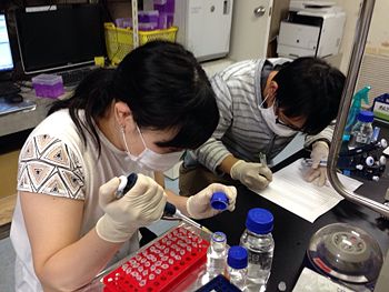
Each staple DNA were dissolved by sterilized water that the concentration of each solution might be 100 μM.

These solutions were taken 2 μL each and mixed.

The volume of the solution was diluted with sterilized water to 500μL. we prepared staple DNA mixture.The sample (50 μM) was prepared mixing M13mp18 (4nM, Takara, Japan), staple DNA mixture (20nM) and 1×TAE/Mg2+ (12.5 mM). This mixture was kept at 90˚C for 10 minutes and cooled from 90˚C to 25˚C at a rate of -1.0˚C/min to anneal the strands.

The annealed mixture (1μL) was deposited on freshly cleaved mica, additional 1X TAE/Mg2+ buffer (40 µL) was added.
We observed this product by AFM. the imaging was performed in the fluid Tapping mode with a BL-AC40TS tip (Olympus, Japan) and performed on a Multimode 8/ Nanoscope system (Bruker AVS).

<html> <font size="4"> <p align="right"><a href="#top">↑ Back to top</a></p> </font> </html>
