Biomod/2014/Kashiwa/Receptor
<html> <head> <div id="#top"></div>
<script type="text/javascript" src="http://code.jquery.com/jquery-1.9.1.min.js"></script>
<script type="text/javascript"> new Image(120,80) = "http://openwetware.org/images/7/7a/Logo2Kashiwa.png";
new image(120,80) = "http://openwetware.org/images/1/1d/Logo2.5.png";
new image(71,143) = "http://openwetware.org/images/2/2a/Top1.gif";
new image(71,143) = "http://openwetware.org/images/f/f0/Top2.gif";
new image() = "http://openwetware.org/images/3/38/BackgroundKashiwa.png";
new image() = "http://openwetware.org/images/9/9c/BackgroundKashiwa2.png"; </script>
<script type="text/javascript">
  $(function(){
     $("#menu li").hover(function(){
        $("ul",this).show();
     },
     function(){
        $("ul",this).hide();
     });
  });
</script>
<script type="text/javascript">
<!--
if (document.images) {
// 設定開始(使用する画像を設定してください)
// 通常の画像 var img0 = new Image(120,80); img0.src = "http://openwetware.org/images/e/ec/LogoKashiwa.png";
// ポイント時の画像1 var img1 = new Image(120,80); img1.src = "http://openwetware.org/images/2/26/Logo3Kashiwa.png";
// 設定終了
} // ポイント時の処理 function On(name) { if (document.images) { document.images['def'].src = eval(name + '.src'); } } // 放した時の処理 function Off() { if (document.images) { document.images['def'].src = img0.src; } } // --> </script>
<META http-equiv="Content-Style-Type" content="text/css">
<style type="text/css">
.page-back {
position: fixed;
bottom: 25px;
right: 25px;}
p.paragraph {
 font-size :100%;
 line-height: 135%;
 margin: 10px 25px;
}
p.reference {
 font-size :90%;
 line-height: 135%;
 margin: 10px 20px;
}
p.indent-paragraph {
 font-size :110%;
 line-height:1.5;
 margin:0 30px;
 text-indent: 1em;
 }
h1.title a{
 font-size :100%;
 display: block;
 text-decoration: none;
 color: #000000;
font-weight:bolder;
 border-left: solid 5px #e00000;
 }
h2.reference a{
 font-size :90%;
 display: block;
 text-decoration: none;
 border-left: solid 5px;
 }
body {
 font-size: 12px;
 font-family: Frutiger, Helvetica, Arial;
 background-color: #B5D3FF;
 overflow-y:scroll;
 overflow-x:hidden;
} article {
background-color: #ffffff
} .container {
  background-color: #ffffff;
  margin-top:0px
}
.OWWNBcpCurrentDateFilled {display: none;}
}
- column-content
{
  width: 0px;
  margin: 0;
  padding: 0;
  float:left;
} .firstHeading {
  display:none;
  width:0px;
}
- globalWrapper
{
  width:900px;
  background-color: #ffffff;
  margin-left: auto;
  margin-right: auto;
}
- column-one
{
  display:none;
  width:0px;
  background-color: #ffffff;
}
- content
{
  margin: 0px;
  align: center;
  padding: 0px 12px;
  width:876px;
  border: 0;
}
- bodyContent
{
  width: 800px;
  padding: 0px 38px;
  align: center;
  background-color: #ffffff;
  position:relative;
}
- column-content
{
  width: 900px;
  background-color: #ffffff;
}
- footer
{
  position: center;
  width: 900px;
}
div.menubar {
  position: fixed;
  background-color: none; /* バーの背景色 */
  opacity: 0.95;
  border-top: 0px double white; /* バーの上端線 */
  border-bottom: 0px double white; /* バーの下端線 */
  min-width: 900px; /* メインメニュー全部が収まる最低横幅 */
  z-index:3
}
div.menubar ul#menu {
  margin: -33px 0px 0px -50px; /* メニューバー外側の余白 */
  padding: 0px; /* メニューバー内側の余白 */
  height: 80px; /* メニューバーの高さ */
  list-style-type: none;
}
div.menubar ul#menu li {
  min-width: 110px; /* メニュー項目の横幅 */
  height: 80px; /* メニュー項目の高さ(「メニューバーの高さ」と一致させる) */
  float: left;
  list-style-type: none;
  list-style-image: none;
  position: relative;
}
div.menubar ul#menu a {
  text-decoration: none; /* メニュー項目の装飾(下線を消す) */
  display: block;
  background-color: #1F003E;/* メニュー項目の背景色 */
  color: white; /* メニュー項目の文字色 */
  line-height: 80px; /* メニュー項目のリンクの高さ(「メニュー項目の高さ」と一致させる) */
  text-align: center; /* メインメニューの文字列の配置(中央寄せ) */
  width: 100%;
  height: 100%;
}
div.menubar ul#menu a:hover {
  background-image: url("http://openwetware.org/images/3/38/BackgroundKashiwa.png"); /* メニュー項目にマウスが載ったときの背景色 */
  color: #8B008B; /* メニュー項目にマウスが載ったときの文字色 */
}
div.menubar ul#menu a:active {
  background-image: url("http://openwetware.org/images/3/33/Background2.png"); /* メニュー項目にマウスが載ったときの背景色 */
  color: #8B008B; /* メニュー項目をクリックした時の文字色 */
}
/* メニューバー直後のClearfix */ div.menubar ul#menu {
zoom:1;
} div.menubar ul#menu:after {
height: 0; visibility: hidden; content: "."; display: block; clear: both;
}
div.menubar ul#menu ul.sub {
  background-color: #1F003E; /* サブメニュー全体の背景色 */
  border-width: 0px 0px 0px 0px; /* サブメニュー全体の枠線の太さ */
  border-style: solid; /* サブメニュー全体の枠線の線種 */
  border-color: #191970; /* サブメニュー全体の枠線の色 */
  margin: 0px;
  padding: 0px;
  display: none;
  position: absolute;
}
div.menubar ul#menu ul.sub li {
  width: 110px;
  height: 50px; /* サブメニュー1項目の高さ */
  border-width: 0px 0px 0px 0px; /* サブメニュー1項目の枠線の太さ */
  border-style: solid; /* サブメニュー1項目の枠線の線種 */
  border-color: #191970; /* サブメニュー1項目の枠線の色 */
  z-index: 3
}
div.menubar ul#menu ul.sub li a {
  line-height: 50px; /* サブメニュー1項目の行の高さ(「サブメニュー1項目の高さ」と合わせる) */
  text-align: center;  /* サブメニュー1項目の項目名の配置(中央寄せ) */
  text-indent: 0px;  /* サブメニュー1項目の項目名前方の余白 */
}
div.menubar ul#menu ul.sub li a:hover {
  background-image: ('back');
  background-size: 100% auto;
  background-color: #FF00FF; /* サブメニュー項目にマウスが載ったときの背景色 */
  color:  #8B008B; /* サブメニュー項目にマウスが載ったときの文字色 */
}
div.menubar ul#menu ul.sub li a:active {
  background-image: ('backclick') /* メニュー項目にマウスが載ったときの背景色 */
  backround-size: 100% auto;
  color: #8B008B; /* メニュー項目をクリックした時の文字色 */
}
</style>
<div class="menubar">
<ul id="menu">
<font face="Frutiger,Helvetica,Arial">
<li><a href="http://openwetware.org/wiki/Biomod/2014/Kashiwa"><img src="http://openwetware.org/images/e/ec/LogoKashiwa.png" onmouseover="this.src='http://openwetware.org/images/7/7a/Logo2Kashiwa.png'" onclick="this.src='http://openwetware.org/images/1/1d/Logo2.5.png'" onmouseout="this.src='http://openwetware.org/images/e/ec/LogoKashiwa.png'" height="80px" width="120px" name="def"></a>
</li>
<li><a href="http://openwetware.org/wiki/Biomod/2014/Kashiwa/Project" onMouseOver="On('img1')" onMouseOut="Off()"><span style="font-size:12pt;">PROJECT</span></a>
<ul class="sub">
<li><a href="http://openwetware.org/wiki/Biomod/2014/Kashiwa/Project#1" onMouseOver="On('img1')" onMouseOut="Off()"><span style="font-size:12pt;">Background</span></a></li>
<li><a href="http://openwetware.org/wiki/Biomod/2014/Kashiwa/Project#2" onMouseOver="On('img1')" onMouseOut="Off()"><span style="font-size:12pt;">Motivation</span></a></li>
<li><a href="http://openwetware.org/wiki/Biomod/2014/Kashiwa/Project#3" onMouseOver="On('img1')" onMouseOut="Off()"><span style="font-size:12pt;">Project Goals</span></a></li>
</ul>
</li>
<li><a href="http://openwetware.org/wiki/Biomod/2014/Kashiwa/Trial" onMouseOver="On('img1')" onMouseOut="Off()"><span style="font-size:12pt;"> EARLY TRIAL </span></a>
<ul class="sub">
<li><a href="http://openwetware.org/wiki/Biomod/2014/Kashiwa/Trial#1" onMouseOver="On('img1')" onMouseOut="Off()"><span style="font-size:12pt;">Design</span></a></li>
<li><a href="http://openwetware.org/wiki/Biomod/2014/Kashiwa/Trial#2" onMouseOver="On('img1')" onMouseOut="Off()"><span style="font-size:12pt;">Approaches</span></a></li>
</ul>
</li>
<li><a href="http://openwetware.org/wiki/Biomod/2014/Kashiwa/Design" onMouseOver="On('img1')" onMouseOut="Off()"><span style="font-size:12pt;">DESIGN</span></a>
<ul class="sub">
<li><a href="http://openwetware.org/wiki/Biomod/2014/Kashiwa/Design#2" onMouseOver="On('img1')" onMouseOut="Off()"><span style="font-size:12pt;">The Receptor</span></a></li>
<li><a href="http://openwetware.org/wiki/Biomod/2014/Kashiwa/Design#1" onMouseOver="On('img1')" onMouseOut="Off()"><span style="font-size:12pt;">The Motor</span></a></li>
</ul>
</li>
<li><a href="http://openwetware.org/wiki/Biomod/2014/Kashiwa/Highlights" onMouseOver="On('img1')" onMouseOut="Off()"><span style="font-size:12pt;"> EXPERIMENT </span></a>
<ul class="sub">
<li><a href="http://openwetware.org/wiki/Biomod/2014/Kashiwa/Highlights" onMouseOver="On('img1')" onMouseOut="Off()"><span style="font-size:12pt;">Highlights</span></a></li>
<li><a href="http://openwetware.org/wiki/Biomod/2014/Kashiwa/Receptor" onMouseOver="On('img1')" onMouseOut="Off()"><span style="font-size:12pt;">The Receptor</span></a></li>
<li><a href="http://openwetware.org/wiki/Biomod/2014/Kashiwa/Motor" onMouseOver="On('img1')" onMouseOut="Off()"><span style="font-size:12pt;">The Motor</span></a></li>
</ul>
<li><a href="http://openwetware.org/wiki/Biomod/2014/Kashiwa/Discussion" onMouseOver="On('img1')" onMouseOut="Off()"><span style="font-size:12pt;"> DISCUSSION </span></a>
<ul class="sub">
<li><a href="http://openwetware.org/wiki/Biomod/2014/Kashiwa/Discussion#1" onMouseOver="On('img1')" onMouseOut="Off()"><span style="font-size:12pt;">Achievements</span></a></li>
<li><a href="http://openwetware.org/wiki/Biomod/2014/Kashiwa/Discussion#2" onMouseOver="On('img1')" onMouseOut="Off()"><span style="font-size:12pt;">Future</span></a></li>
</ul>
</li>
<li><a href="http://openwetware.org/wiki/Biomod/2014/Kashiwa/Protocols" onMouseOver="On('img1')" onMouseOut="Off()"><span style="font-size:12pt;">PROTOCOL</span></a>
</li>
<li><a href="http://openwetware.org/wiki/Biomod/2014/Kashiwa/Team" onMouseOver="On('img1')" onMouseOut="Off()"><span style="font-size:12pt;">TEAM</span></a>
<ul class="sub">
<li><a href="http://openwetware.org/wiki/Biomod/2014/Kashiwa/Team#1" onMouseOver="On('img1')" onMouseOut="Off()"><span style="font-size:12pt;">Members</span></a></li>
<li><a href="http://openwetware.org/wiki/Biomod/2014/Kashiwa/Team#2" onMouseOver="On('img1')" onMouseOut="Off()"><span style="font-size:12pt;">Sponsors</span></a></li>
</ul>
</li>
</font>
</ul>
</div> <br> <br> <br> <div class="page-back"><a href="#top"> <img src="http://openwetware.org/images/2/2a/Top1.gif" onmouseover="this.src='http://openwetware.org/images/f/f0/Top2.gif'" onmouseout="this.src='http://openwetware.org/images/2/2a/Top1.gif'" height="150px"/></a></div> </head>
</html>
<html>
<head> <style>
div.imagebox {
  border: 1.7px dashed #2E2EFE; /* 1.枠線 */
  background-color: #F0F8FF;  /* 2.背景色 */
  width: 290px; /* 横幅 */
  float: right; /* 右に配置 */
  margin: 0px 30px 0px 10px;
} p.image {
  text-align: center;
  width: 280px;
  height: 173px;
  margin: 5px;
} p.caption {
  margin: 5px 10px;
  font-size: 90%;  /* 5.文字サイズ */
  color: black; /* 6.文字色 */
}
div.centerbox {
  border: 1.7px dashed #2E2EFE; /* 1.枠線 */
  background-color: #F0F8FF;  /* 2.背景色 */
  height:auto;
  width: 290px; /* 横幅 */
  margin: 0px 40px;
  float: left;
}
div.yokobox{
  border: 1.7px dashed #2E2EFE; /* 1.枠線 */
  background-color: #F0F8FF;  /* 2.背景色 */
  height:150px;
  width: 310px; /* 横幅 */
  margin: 0px 40px;
  float: right;
}
p.yokoimage{
  text-align: center;
  width: 300px;
  height: 120px;
  margin: 5px;}
div.autobox {
  border: 1.7px dashed #2E2EFE; /* 1.枠線 */
  background-color: #F0F8FF;  /* 2.背景色 */
  height:auto;
  width: auto; /* 横幅 */
  margin: 5px;
  float: right;
}
div.smallbox {
  border: 1.7px dashed #2E2EFE; /* 1.枠線 */
  background-color: #F0F8FF;  /* 2.背景色 */
  width: 210px; /* 横幅 */
  margin: 5px;
  float: right;
} p.smallimage {
  text-align: center;
  width: 200px;
  height: 120px;
  margin: 5px;
}
h1.big{
 font-size :16px;
 text-decoration: none;
 color: #000000;
 margin: 10px 15px;
 font-weight: bolder;
 }
h1.sub {
 font-size:20px;
 font-weight: bolder;
 margin: 10px 5px;
}
p.menu {
 font-size :100%;
 line-height: 135%;
 margin: 0px 40px;
} </style>
</head>
<body>
EXPERIMENTS
<a name="background"> 1. The Sensing System: The Receptor</a>
<img src="http://openwetware.org/images/c/c3/Receptorexperiments.png" height="248px" weight="300px" align="right">
This section described the details of our approach to develop the sensing system "Receptor" of PoLICe. The approach mainly consists of four steps as follows.
- <a href="#1-1">1-1. Preparation of the components </a>
- <a href="#1-1-1">1-1(a). Folding of the Wall </a>
- <a href="#1-1-2">1-1(b). Production of MISTIC </a>
- <a href="#1-1-3">1-1(c). Design of the Activator </a>
- <a href="#1-2">1-2. Embedment of the Wall into the liposome </a>
- <a href="#1-3">1-3. Linkage of the Activator to the liposome </a> <a name="1-1"></a>
- <a href="#1-4">1-4. Control of the Wall conformation</a>
<a name="1-1-1"></a>
1-1. Preparation of the components
Three components were prepared to develop the Receptor: The Wall of DNA origami, MISTIC and the Activator. Please check our <a href="http://openwetware.org/wiki/Biomod/2014/Kashiwa/Design">Design page</a> to check each component's role.
1-1(a). Folding of the Wall
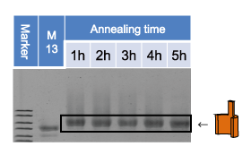
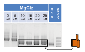
| <img src="http://openwetware.org/images/9/99/Walltimeaneal.png" width="180px"> | <img src="http://openwetware.org/images/2/29/Annealringase.png" width="180px"> | <img src="http://openwetware.org/images/1/1c/Wallnaclkashiwa.png" width="180px"> | <img src="http://openwetware.org/images/e/e0/Wallmgcl2l2.png" width="180px"> |
|---|---|---|---|
Fig.1-1(a)-1. Gel analysis of the Walls annealed in different time. | Fig.1-1(a)-2. Gel analysis of the Walls annealed in different temperature. | Fig.1-1(a)-3. Gel analysis of the Walls annealed in different concentration of NaCl. | Fig.1-1(a)-4. Gel analysis of the Walls annealed in different concentration of MgCl2. |
In this experiment, the assembly condition of the Wall structure was optimized and results were analyzed by agarose gel electrophoresis. The optimum conditions were confirmed by comparing migration distances of each samples. The sample of which migration distance is the longest was regarded as the optimum condition.
| <IMG src="http://openwetware.org/images/c/c9/Wall_temforwiki.JPG" width="300"/> |
Fig.1-1(a)-5. TEM image for the Motor-Monomers. |
The optimum results are as follows.
- Concentration of MgCl2 : 15 mM
- Temperature of annealing : 45.3 °C
- Time of annealing : 5 hours
- Concentration of NaCl : 2.5 mM
<a name="1-1-2"></a>
The folding is confound by the TEM image (Fig.1-1(a)-5.).
1-1(b). Production of MISTIC
(i) Mistic gene mutation
We introduced the mutation to the MISTIC, a protein which penetrates the membrane for 4 times, by Quick Change method. We introduce Cys residue at the position after first α-herix for connection with enzyme or fluorescent labeling (29th valine to cysteine).
The wild type of the configuration of MISTIC was as follows.
MFCTFFEKHHRKWDILLEKSTGVMEAMKVTSEEKEQLSTAIDRMNEGLDAFIQLYNESEIDEPLIQ LDDDTAELMKQARDMYGQEKLNEKLNTIIKQILSISVSEEGEKE
- We made 4 types of mutants (Cys3 to Val, Val29 to Cys, Cys between 84-85 and Cys between 110-111), but only used this one.
- Underlined parts form α-helix structures which are considered to be the membranes penetrating domain.
- A red letter shows the part which we introduced variation in this experiment.
We confirmed the introduction of mutation by sequencing.
(ii) Preparation of templateDNA of MISTIC for PUREfrex
<img src="http://openwetware.org/images/d/dd/Izuta1.png" width="280px" height="173px">
Fig.1-1(b)-1. Agarose gel electrophoresis of PCR products.
In order to express MISTIC by PURE(protein synthesis using recombinant elements)frex, we added T7 promoter, SD sequence and T7 terminator to the mutatn DNA. At the same time, we substituted valine for Cys3 then completed to make mono-cysteine mutant. We also added Histag to the C-terminus and replaced some amino acids near N-terminus by amber, one of a termination codon, to introduce biotin to the N-terminus using unnatural amino acids.
We tried first seven amino acids of Mistic for biotin insertion, and found that position 3 is the best place.
M+VTFFEKHHRKWDILLEKSTGVMEAMKCTSEEKEQLSTAIDRMNEGLDAFIQLYNESEIDEPLIQLDDD TAELMKQARDMYGQEKLNEKLNTIIKQILSISVSEEGEKEHHHHHH
- + is an abbreviation for unnatural amino acid.
- PURE frex is reconstructed cell-free system for transcription and translation reaction.
We checked the PCR product by agarose electrophoresis, and conformed that there are no extra band and the length is that of expected one (455bp).
(iii) Expression and purification of MISTIC protein
1. Expression
<img src="http://openwetware.org/images/9/99/Izuta2.png" width="280px">
Fig.1-1(b)-2. SDS-PAGE of PURE products.
We added MISTIC gene to reaction solution, incubated at 37℃ for 2 hours and obtained MISTIC protein. The expression in PUREfrex was confirmed by SDS-PAGE. In order to distinguish between the factors of PUREfrex and MISTIC, we added 35S-Met to PUREfrex reacton solution, labelled MISTIC with a radioisotope and detected it by Photostimulated luminescence (PSL: BAS).
As we could observe the band at the aimed position (13.6kDa), we considered that MISTIC protein was expressed correctly in PURE frex.
2. Purification
<img src="http://openwetware.org/images/0/06/Izuta3.png" width="280px";>
Fig.1-1(b)-3. SDS-PAGE of PURE products and Purified Products.
We purified products of PUREfrex using Histag columnand by SDS-PAGE.
As we could see MISTIC in the elusion fraction and impurities were few, we concluded that the purification was succeeded.
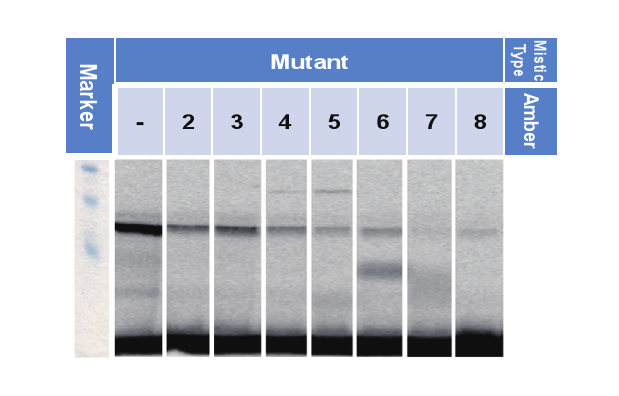
(ⅳ) Confirmation of the introduction of unnatural amino acids
<img src="http://openwetware.org/images/0/03/Izuta-4.png" width="300px">
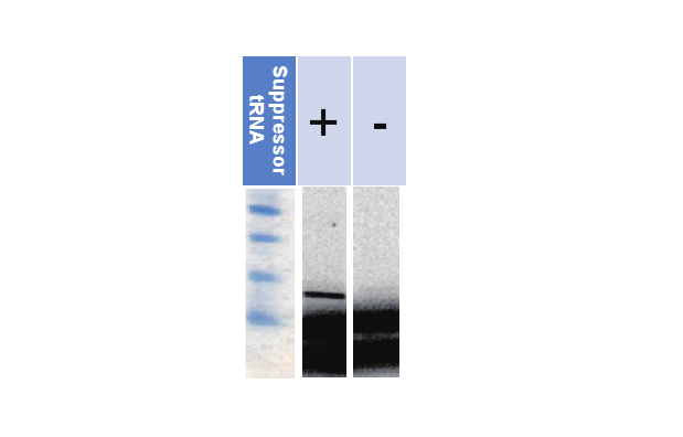
Fig.1-1(b)-4. SDS-PAGE of PURE products
We would assay that that unnatural amino acids are included in MISTIC, by confirming that MISTIC expresses with suppresser tRNA and but not without the suppressor tRNA. We detected MISTIC labelled by a radioisotope by PSL (same as the method explained in “3.Expresion”).
As we could see that MISTIC is not expressed when unnatural amino acids are not added, we could determine that tRNA which sets into amber competitively with RF1 exists little. From this result, we could confirm that unnatural amino acids are included in MISTIC.
(v) Confirmation of the ability of access to biotin under the non-denatured condition
| <img src="http://openwetware.org/images/f/ff/Izuta-5.png" width="240px"> |
|---|
Fig.1-1(b)-5. SDS-PAGE of PURE products |
We confirmed accessibility of streptavidin(SA) to biotin under the non-denatured condition by certifying whether or not MISTIC are removed by the magnetic beads which is coupled to SA. To exclude the influence of other PURE factors, we labelled Mistic by 35S-Met and assayed by PSL (this method is mentioned above).
We loaded same amount of the product of PURE to all lanes. Lane shows that the amount of Mistic with biotin is decreased; indicating that the introduced biotins are accessible by SA.
(vi) Confirmation of Cysteine residue modification of MISTIC to cysteine
We labelled MISTIC by Cy5 maleimide (Cy5-MA) and confirmed whether cysteine residue of MISTIC is able to be modified or not.
We mixed 0.3µL of 10mM Cy5-MA and 50µL of 3.26µM MISTIC and incubated for 30 minutes. Products were conformed by SDS-PAGE after G-50 column purification and Cy5 fluorescence was observed. After that, protein was stained by SYPRO ORANGE and fluorescence was observed again.
<a name="1-1-3"></a>
As we could see the fluorescence of Cy5 at the position of the band of MISTIC, the modification seemed to be successful. Comparing the intensity of fluorescence with other lanes which we put the same amount of Cy5-MA, the modification rate is presumed to be --%.
1-1(c). Design of the Activator
Two restriction enzymes, HindⅢ and Lambda Exonuclease, were compared to choose which is appropriate for the Activator. Two aspects were evaluated in this experiment: modification with oligonucleotides and enzyme activity.
(i) Evaluation of modification with oligonucleatides
<img src="http://openwetware.org/images/a/a5/BS%28PEG%299.png" width="280px">
Fig.1-1(c)-1. BS(PEG)9 for oligo-modification.
HindⅢ and Lambda Exonuclease are modified with oligonucleotides to join the Activator and the Anchor. BS(PEG)9 was used for the modification.
On HindⅢ, the modification was analyzed by Native-PAGE. Oligonucleotides were labelled with Cy3 and HindⅢ was stained with SYPRO Orange to confirm the modification.
<img src="http://openwetware.org/images/3/30/Atuta2.png">
Fig.1-1(c)-2. Cy3 fluorescence image of oligo-modified HindⅢ by Native-PAGE.
<img src="http://openwetware.org/images/a/ae/Atuta3.png">
Fig.1-1(c)-3. SYPRO-ORANGE fluorescence image of oligo-modified HindⅢ by Native-PAGE.
In the pictures observed with both of cy3 and SYPRO-Orange, a band was observed at the same position in lane 3. Therefore, it was confirmed that Hind3 was labelled with cy3-oligonucleotide. (Multiple band of Hind3 was from the first.)
On Lamda Exonuclease, the modification was confirmed by Native-PAGE in the same way as HindⅢ.
<img src="http://openwetware.org/images/b/b1/Atuta4.png">
Fig.1-1(c)-4. Cy3 fluorescence image of oligo-modified Lamda Exonuclease by Native-PAGE.
<img src="http://openwetware.org/images/7/73/Atuta5.png">
Fig.1-1(c)-5. SYPRO-ORANGE fluorescence image of oligo-modified Lamda Exonuclease by Native-PAGE.
The result shows Lamda Exonuclease was modified with oligonucleotide successfully.
(ii) Evaluation of enzyme activity
| <img src="http://openwetware.org/images/6/67/Lambda_Exonuclease_reaction.png" width="280px"> | <img src="http://openwetware.org/images/8/86/Hind3_reactionKashiwa.png" width="280px"> |
|---|---|
| <img src="http://openwetware.org/images/2/2e/Hindmodify.png" width="280px"> | <img src="http://openwetware.org/images/7/78/1-1%28c%29-7-C.png" width="280px"> |
Fig.1-1(c)-7. DNA cleavage reaction by HindⅢ. |
Fig.1-1(c)-8. DNA cleavage reaction by Lamda Exonuclease. |
HindⅢ is an restriction endonuclease that recognizes base sequence 5’-AAGCTT-3’ and cleaves it, while Lambda Exonuclease degrades one strand from 5'-phosphoryl termini of dsDNA.
In this experiment, enzyme activity of HindⅢ and Lambda Exonuclease was evaluated in various conditions. The purpose is to confirm that HindⅢ and Lambda Exonuclease have activity in condition for DNA origami. The activity was analyzed by Native-PAGE.
<img src="http://openwetware.org/images/4/40/1-1%28c%29-9.png">
Fig.1-1(c)-9. Native-PAGE shows Lamda activity depending on PH.
<img src="http://openwetware.org/images/6/6a/1-1%28c%29-8.png">
Fig.1-1(c)-8. Native-PAGE shows HindⅢ activity depending on PH.
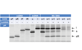
First, enzyme activity depending on PH was analyzed.
The result showed that Lambda Exonuclease can act in pH7.5-8.5. In pH9.0 and pH9.4, dsDNA seems to be unstable. So, pH7.5-8.5 is appropriate for reaction of Lambda Exonuclease and DNA.
<img src="http://openwetware.org/images/7/71/1-1%28c%29-11.png">
Fig.1-1(c)-11. Native-PAGE shows Lamda activity depending on concentration of MgCl2.
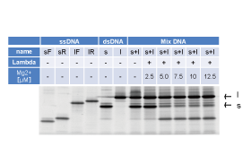
Then enzyme activity depending on concentration of MgCl2 was analyzed.
The result showed that Lambda Exonuclease can act in 10mM MgCl2.
<a name="1-2"></a>
(i) Evaluation of required distance between DNA origami and recognition-sequence
In this experiment, how much distance between DNA origami and recognition-sequence is required to work enzyme was evaluated. If the substrate of enzyme is too close to other structure, enzyme may not be able to access to it.
We used barrel-shaped DNA origami as an obstacle and substrate dsDNA was attached to it. Whether the enzyme can access to substrate was analyzed with Native-PAGE.
・HindⅢ
Method
1) Annealed barrel structure with substrate of enzyme.
2) Reacted enzyme with barrel.
3) Analyzed by Native-PAGE
4) Observed with Cy3
5) Stained the gel by SYBR-Gold.
6) Observed by the LAS-4000
Result
HindⅢ had an activity in all conditions, and when recognition-sequence was located in a position 11 bases from barrel, HindⅢ had the highest activity.
・Lambda Exonuclease It’s reaction product is ssDNA and nucleotides. So, it’s difficult to detect whether it acts to substrate and we couldn’t evaluate the access range of Lambda Exonuclease.
Since then, HindⅢ was adopted as the Activator.
1-2. Embedment of the Wall into the liposome
Embedding the Wall into the liposome needs two steps: modifying the Wall with cholesterol and putting them into the liposome. The experiments were done individually.
(i) Hybridization of cholesterol oligomer with the Walls
In this experiment, we examined the best concentration and the number of staples which the cholesterol oligomers can connect. Hybridization of cholesterol oligomer with the Walls is needed to penetrate the Walls into the membrane of liposomes. The result was assayed by 1% agarose gel electrophoresis.
If the cholesterol oligomer was connected to the Walls the band would be smear. In sample 2_1~4, 3_1, 3_2, 5_1, 5_2, 6_1. Considering that sample 2 and 3 do not have staple for hybridization, the Walls of sample 2 and 3 aggregated by the existence of cholesterol oligomer. Sample 5_1, 5_2 and 6_1 are considered to be successful in hybridization. As the band of sample 5_2 and 6_1 are weaker than that of sample 5_1, we decided that the best condition was sample5_1.
(ii) Putting the Monomers into the liposome
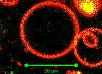
In this experiment, we tried to put the Walls into giant unilamellar vesicles (GUV) and penetrate them into the inner membrane of GUV. The biotin labeled Walls were combined with streptavidin coated Q-dot and were put into GUV. Inclusion of the Walls was confirmed by confocal microscope. The number of GUV was estimated by measuring the fluorescence intensity of Nile Red using Plate reader (Wallac 1420 MULTILABEL COUNTER, Perkin Elmer life sciences). We estimated that the density of lipid in GUV solution was 0.2~0.7mg/mL. Supposing that the diameter of GUV is 10µm, we calculated that there are 2×10¹¹~6×10¹¹ vesicles/L. As we got 150µL of GUV solution per sample, meaning that there are 3×10⁷~9×10⁷ vesicles per sample.
In sample1 (GUV including cholesterol hybridized Walls), the density of the Walls was 16nM. Considering that we put 2.5µL of the Walls in the inner solution of GUV, there should be 2.4×10¹⁰ Walls in sample1. From these assumption, we calculated that 267~800 Walls might be included in single GUV. In the same way, considering that the density of the Walls in sample2 was 26nM, there might be 434~1300 Walls/GUV in sample2 and 333~100 Qdot/GUV in sample3.
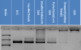
The images observed by confocal microsocope are shown below. The connection of the Walls and Qdot was assayed by 1% agarose gel electrophoresis.
The images observed by confocal microsocope are shown below.
The connection of the Walls and Q-dot was assayed by 1% agarose gel electrophoresis.
1) Image of confocal microscope
| <img src="http://openwetware.org/images/e/e8/Yui1t.png" width="180px"> | <img src="http://openwetware.org/images/e/ed/Yui2t.png"width="180px"> | <img src="http://openwetware.org/images/0/0b/Hqdotkashiwa.png"width="180px"> | <img src="http://openwetware.org/images/7/7d/Hyuyuikashiwa.png"width="180px"> |
|---|---|---|---|
| Fig.1-2-1. | Fig.1-2-2. | Fig.1-2-3. | Fig.1-2-4. |
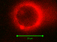
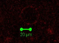
GUVs were stained by Nile Red. Yellow green dots indicate the fluorescence of Nile Red and red dots indicate the fluorescence of Qdot-Walls.
Comparing the four pictures, we did not observe red dots inside GUV only sample in Fig. 4. From this fact, we concluded the inclusion of the Walls and Qdot into the GUV. However, as we could see the red dots sticking to the membrane of GUV in sample of cholesterol-less Walls, we could not prove that the Walls penetrated into GUV using the support of cholesterol, but Walls or Qdot itself might have the potential to insert or attach to the GUV membrane. Also, as the excitation and fluorescence emission wave length of Nile Red and Qdot were close, the membrane of GUV was shown in red even in the GUV image not including the Qdot. To prevent this, we tried different Qdot having peak fluorescent emission peak far from that of Nile Red, but similar thing happened. We also tried to observe GUV without Nile Red staining, but fail to prevent the non-specific binding of Qdot or cholesterol-less Wall, suggesting that the key of this kind of experiments were preventing such non-specific binding.
2) Agarose gel electrophoresis
| <img src="http://openwetware.org/images/e/e4/Yui3443kashiwsa.png" width="250px"> | <img src="http://openwetware.org/images/8/81/Fig.1-2-6._Agarose_gel_electrophoresis_showing_the_existence_of_DNA.png" width="250px"> | <img src="http://openwetware.org/images/2/20/Fig.1-2-7._Merged_image_of_fig._1-2-5%2C_fig.1-2-6.png" width="250px"> |
|---|---|---|
| Fig.1-2-5. | Fig.1-2-6. | Fig. 1-2-7. |
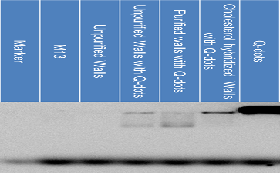
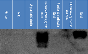
Fig.1-2-5 shows the existence of Qdot and fig.1-2-6. shows the existence of DNA. Fig.1-2-7 shows the merged image of the two. Comparing the fifth, sixth and seventh lane from the left in fig. 1-2-5, we can see the difference in the position of the Qdot. In the second picture, as the band of the sixth lane from the left is smear, we could say that cholesterol oligomers hybridized to the Walls. In the third picture, as we could see the band of the fifth and sixth lane from the left are at the same position, we could conclude that Qdot connected to the Walls. This data shows that the Qdot connected to the Walls through the connection of biotin and streptavidin.
As we could not observe the Walls penetrated into GUV from inside of it, we tried to penetrate them from outside of it in the next experiment. The number of the Walls penetrated into GUV was estimated in the same way as we have explained before. In this experiment, we put 2.5µL of Walls to 12.5µL of GUV. We calculated that there should be 4000~12000 Walls/GUV in sample1 and 3, 5000~15000 Walls/GUV in sample2. The Walls in sample1 (The Walls(i)) have biotin staple, and the Walls in sample3 (The Walls(iii)) do not have biotin staples. We observed the mixture of GUV and the Walls by confocal microscope and 1% agarose gel electrophoresis. The images observed in the microscope are shown below.
1) Image of confocal microscope
| <img src="http://openwetware.org/images/2/20/Fig.1-2-8._Confocal_microscope_image_of_GUV_including_cholesterol_hybridized_Walls%28i%29_with_Qdot.png" width="250px"> | <img src="http://openwetware.org/images/5/55/Fig.1-2-9._Confocal_microscope_image_of_GUV_including_non-cholesterol_hybridized_Walls%28i%29_with_Qdot.png" width="250px"> | <img src="http://openwetware.org/images/e/e5/Fig.1-2-10._Confocal_microscope_image_of_GUV_including_non-cholesterol_hybridized_Walls%28iii%29_with_Qdot.png" width="250px"> |
|---|---|---|
| Fig. 1-2-8. | Fig. 1-2-9. | Fig.1-2-10. |
The second and the third picture were observed using the same observation condition (intensity of the laser, size of pinhole, gain of the detector). The first picture was observed using the weaker laser.
Comparing the first and the third pictures, we could see that the intensity of the red dots is much weaker in the third picture even the laser was stronger in the third picture. As the Walls used in the sample of third picture do not have biotin for cholesterol oligomers to hybridize, Qdot almost disappeared after the purification. Some of the Qdot seemed to have stuck to the lipid of GUV, so the weak fluorescence of Qdot was observed. In the second picture, the red dots around the GUV is smear, but almost all of the dots seen in the picture are quite near the GUV, so the Walls seemed to be stuck to the GUV. So again, we have to think of the way to prevent the Walls stick to GUV non-specifically and that would be our future work.
2) Agarose gel electrophoresis
| <img src="http://openwetware.org/images/e/e5/Fig.1-2-11.png" width="250px"> | <img src="http://openwetware.org/images/3/3a/Fig.1-2-12.png" width="250px"> | <img src="http://openwetware.org/images/4/40/Fig.1-2-13.png" width="250px"> |
|---|---|---|
| Fig. 1-2-11. | Fig. 1-2-12. | Fig.1-2-13. |
Fig.1-2-11 shows the existence of Qdot and fig.1-2-12 shows the existence of DNA. Fig.1-2-13 shows the merged image of the two. The sixth lane from the left in the second picture suggests that the Walls(i) was cholesterol hybridized because the position of the band changed from that of the Walls(i) not including cholesterol oligomers.
The sixth lane from the left in the third picture show that the band of the Qdot and the Walls(i) appeared at the same position, so we could say that the connection of Qdot-streptavidin and the Walls(i) was successful.
| <img src="http://openwetware.org/images/b/b1/Fig.1-2-14.png"> | <img src="http://openwetware.org/images/b/b1/Fig.1-2-14.png"> |
|---|---|
| Fig. 1-2-14. | Fig. 1-2-15. |
Fig.1-2-14 shows the existence of Qdot and fig.1-2-15 shows the existence of DNA.
We can see the band in the sixth lane from the left in the first picture, but we cannot see any band in the same lane in the second picture. This means that Qdot did not stick to the Walls(iii). In the image of the confocal microscope, we could see some Qdot sticking to GUV, so the amount of the Qdot remained after purification was too small to be observed by electrophoresis but large enough to be observed by the confocal microscope.
<a name="1-3"></a>
1-3. Linkage of the Activator to the liposome
<a name="1-4"></a>
1-4. Control of the Wall conformation
Wall and Mistic form the complex through avidin-biotin binding using divalent SA on the membrane and biotins on each. On Mistic’s side, biotin was integrated into the polypeptide of Mistic itself using suppressor tRNA in translation. On Wall’s side, biotin is connected to Wall with dsDNA linker (22 bp) which contains restriction endonuclease recognition site.
The sequence of linker is as follows.
ssDNA1:
ATGGATGGTAAGCTTCTTCTCGtttttttTTAATATATGTGAGTGTTAATTAGGGG GAGGCGGTT
ssDNA2:
[Cy3]-CGAGAAGAAGCTTACCATCCATtttttttttttttttttttt-[Bio]
- underlined part are annealed into Wall and, red letters indicate recognition site of Hind3.
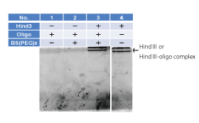
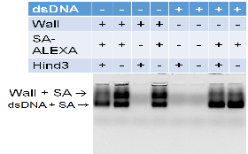
Formation and decomposition of Wall- SA complex in solution
We mixed Purified Wall and SA-Alexa647 and incubated for 1h at room temperature. Then we added Hind3 and incubated for 1h again at room temperature.
The formation and decomposition was conformed by agarose electrophoresis and Cy3, Cy5 and EtBr fluorescent were observed.
In the gel images, Cy3 signal indicates the position of Wall before Hind3 treatment, but low melting temperature of the Hind3 cleaved product induce the release of Cy3 containing fragment, therefore after Hind3 treatment, the Cy3 signal indicate merely the cleaved dsDNA fragment. Also Alexa647 and EtBr signal indicates the position of SA and DNA origami, respectively.
| <img src="http://openwetware.org/images/3/32/1-4_cy3.png" width="250px"> | <img src="http://openwetware.org/images/f/fc/1-4_cy5.png" width="250px"> | <img src="http://openwetware.org/images/5/5a/1-4_etbr.png" width="250px"> |
| Cy3 labeled. | Cy5 labeled. | EtBr labeled. |
|---|
In lane 2 new band containing both Cy3 (biotin-oligonucleotide) and Alexa647 (SA) signal was observed, indicating the formation of the Wall-SA complex.
Besides that, we conformed the Hind3 induced decomposition of Wall-SA complex by the diminishing of complex band in Cy3 and Alexa647.
<a href="http://openwetware.org/wiki/Biomod/2014/Kashiwa/Motor"><img src="http://openwetware.org/images/a/a8/Monomerswitch.png" onmouseover="this.src='http://openwetware.org/images/0/04/Monomerswitch2.png'" onmouseout="this.src='http://openwetware.org/images/a/a8/Monomerswitch.png'" height="160px" width="240px" style="padding:5px 60px;"></a>
Reference
1. Science. 2005 Feb 25;307(5713):1317-21.
NMR structure of Mistic, a membrane-integrating protein for membrane protein expression.
Roosild TP1, Greenwald J, Vega M, Castronovo S, Riek R, Choe S.
2. Angew Chem Int Ed Engl. 2014 Jul 14;53(29):7535-8. doi: 10.1002/anie.201403929. Epub 2014 Jun 4.
In vitro synthesis of the E. coli Sec translocon from DNA.
Matsubayashi H1, Kuruma Y, Ueda T.
3. Proc Natl Acad Sci U S A. 2013 Apr 30;110(18):7276-81. doi: 10.1073/pnas.1303857110. Epub 2013 Apr 15.
Detergent-mediated incorporation of transmembrane proteins in giant unilamellar vesicles with controlled physiological contents.
Dezi M1, Di Cicco A, Bassereau P, Lévy D.
</body>
<footer style="position:relative; left:600px;"> © 2014 UTokyo Chem & Bio </footer>