Biomod/2014/Kashiwa/Motor: Difference between revisions
Saki Atsuta (talk | contribs) No edit summary |
No edit summary |
||
| (44 intermediate revisions by 2 users not shown) | |||
| Line 3: | Line 3: | ||
<html> | <html> | ||
<head> | <head>- | ||
<style> | <style> | ||
| Line 91: | Line 91: | ||
<li> <a href="#2-1-3"> 2-1(c). Equipment of the Motor-Monomer with divalent SA </a> | <li> <a href="#2-1-3"> 2-1(c). Equipment of the Motor-Monomer with divalent SA </a> | ||
</ul> | </ul> | ||
</li><a name="#2-1"></a> | </li><a name="#2-1"></a><a name="2-1"></a> | ||
<li> <a href="#2-2">2-2. Deactivation and reactivation of the binding activity of streptavidin</a></li | <li> <a href="#2-2">2-2. Deactivation and reactivation of the binding activity of streptavidin</a></li> | ||
<li> <a href="#2-3">2-3. Incorporation of the Motor-Monomers into the liposome </a></li> | <li> <a href="#2-3">2-3. Incorporation of the Motor-Monomers into the liposome </a></li><a name="2-1-1"></a> | ||
</ul> | </ul> | ||
<br clear="right"> | <br clear="right"> | ||
<h1 class="sub">2-1. Production of the Motor-Monomer </h1 | <h1 class="sub">2-1. Production of the Motor-Monomer </h1> | ||
<p class="paragraph"> | <p class="paragraph"> | ||
| Line 122: | Line 122: | ||
</TR> | </TR> | ||
<TR> | <TR> | ||
<TD><p class="cap">Fig.2-1(a)-5. TEM image for the | <TD><p class="cap">Fig.2-1(a)-5. TEM image for the Motor-Monomer.</p></TD> | ||
</TR> | </TR> | ||
</TABLE> | </TABLE> | ||
| Line 140: | Line 140: | ||
<br> | <br> | ||
<p class="paragraph"> | <p class="paragraph"><a name="2-1-2"></a> | ||
The folding is corroborated by the TEM image (in fig.2-1(a)-5.) | The folding is corroborated by the TEM image (in fig.2-1(a)-5.) | ||
</p> | </p> | ||
<br clear="right"> | <br clear="right"> | ||
<h1 class="big">2-1(b). Synthesis of divalent streptavidin</h1> | <h1 class="big">2-1(b). Synthesis of divalent streptavidin</h1> | ||
<div class="imagebox"> | <div class="imagebox"> | ||
<p class="image"><img src=""></p> | <p class="image"><img src="http://openwetware.org/images/b/ba/2-1%28b%29-1.png"></p> | ||
<p class="caption">Fig.2-1(b)-1. SDS-PAGE showing purified divalent SA.</p> | <p class="caption">Fig.2-1(b)-1. SDS-PAGE showing purified divalent SA.</p> | ||
</div> | </div><br> | ||
<p class="paragraph"> | <p class="paragraph"> | ||
In this experiment, divalent streptavidin (SA) was synthesized for equipment to the Motor-Monomers. | In this experiment, divalent streptavidin (SA) was synthesized for equipment to the Motor-Monomers. | ||
| Line 161: | Line 165: | ||
<div class="imagebox"> | <div class="imagebox"> | ||
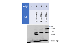
<p class="image"><img src=""></p> | <p class="image"><img src="http://openwetware.org/images/2/28/2-1%28b%29-2.png"></p> | ||
<p class="caption">Fig.2-1(b)-2. SDS-PAGE showing the affinity of divalent SA with biotin-modified oligos.</p> | <p class="caption">Fig.2-1(b)-2. SDS-PAGE showing the affinity of divalent SA with biotin-modified oligos.</p> | ||
</div> | </div><br> | ||
< | |||
<p class="paragraph">The affinity of divalent SA with biotin-modified oligonucleotides was then analyzed by SDS-PAGE.</p> | |||
<a name="2-1-3"></a> | |||
<br clear="right"> | <br clear="right"> | ||
| Line 181: | Line 185: | ||
<center> | <center> | ||
<img src="http://openwetware.org/images/7/72/Click1.png" width="700px"> | |||
<p class="cap">Fig.2-1(c)-1. Optimum combinations of click reaction.</p> | <p class="cap">Fig.2-1(c)-1. Optimum combinations of click reaction.</p> | ||
</center> | </center> | ||
| Line 194: | Line 192: | ||
First, the optimum combination of click reaction between simple alkyne and azide was analyzed. two pairs out of 16 pairs were consequently chosen as the optimum combinations.</p> | First, the optimum combination of click reaction between simple alkyne and azide was analyzed. two pairs out of 16 pairs were consequently chosen as the optimum combinations.</p> | ||
<div class="centerbox"> | |||
<p class="image"><img src="http://openwetware.org/images/a/a7/2-1%28c%2922.png"></p> | |||
<p class="caption">Fig.2-1(c)-2. Analysis of click reaction by Native-PAGE (16 pairs.)</p> | |||
Fig.2-1(c)-2. Analysis of click reaction by Native-PAGE (16 pairs.) | </div><br> | ||
<div class="centerbox"> | |||
Fig.2-1(c)-3. Analysis of click reaction by Native-PAGE (4 pairs.) | <p class="image"><img src="http://openwetware.org/images/1/16/2-1%28c%2932.png"></p> | ||
<p class="caption">Fig.2-1(c)-3. Analysis of click reaction by Native-PAGE (4 pairs.)</p> | |||
Fig.2-1(c)-4. Analysis of click reaction by Native-PAGE (2 pairs.) | </div><br> | ||
<div class="centerbox"> | |||
<p class="image"><img src="http://openwetware.org/images/f/f9/2-1%28c%2942.png"></p> | |||
<div class=" | <p class="caption">Fig.2-1(c)-4. Analysis of click reaction by Native-PAGE (2 pairs.)</p> | ||
</div><br> | |||
<div class="centerbox"> | |||
<p class="image"><img src="http://openwetware.org/images/5/55/Table2-1-c-1.png" width=“160px” height="100px"></p> | |||
<p class="caption">Table 2-1(c)-1</p> | |||
</div><br> | |||
<div class="centerbox"> | |||
<p class="image"><img src="http://openwetware.org/images/e/ea/Gonza_click.png"></p> | <p class="image"><img src="http://openwetware.org/images/e/ea/Gonza_click.png"></p> | ||
<p class="caption">Fig.2-1(c)-5.Relation between time and rate of reaction | <p class="caption">Fig.2-1(c)-5.Relation between time and rate of reaction</p> | ||
</div> | </div><br> | ||
< | |||
<p class="paragraph">In this experiment, as the reaction was regarded as pseudo-first-order reaction, trendline was fitted and kon was gotten. There were few difference between 2pairs in reactivity.</p> | <p class="paragraph">In this experiment, as the reaction was regarded as pseudo-first-order reaction, trendline was fitted and kon was gotten. There were few difference between 2pairs in reactivity.</p> | ||
| Line 223: | Line 219: | ||
<p class="paragraph"><b>(ii) Evaluation of biotin-binding activity of divalent SA</b></p> | <p class="paragraph"><b>(ii) Evaluation of biotin-binding activity of divalent SA</b></p> | ||
Fig.2-1(c)-6. Analysis of NHS reaction by Native-PAGE (Cy5). | <div class="centerbox"> | ||
Fig.2-1(c)-7. Analysis of NHS reaction by Native-PAGE (SYBR Gold). | <p class="image"><img src="http://openwetware.org/images/8/8b/2-1%28c%2962.png"></p> | ||
<p class="caption">Fig.2-1(c)-6. Analysis of NHS reaction by Native-PAGE (Cy5).</p> | |||
</div><br> | |||
<div class="centerbox"> | |||
<p class="image"><img src="http://openwetware.org/images/c/c8/2-1%28c%2972.png"></p> | |||
<p class="caption">Fig.2-1(c)-7. Analysis of NHS reaction by Native-PAGE (SYBR Gold).</p> | |||
</div><br> | |||
<p class="paragraph">Figure 2-1(c)-1 shows that SA was linked with | <p class="paragraph">Figure 2-1(c)-1 shows that SA was linked with Cy5-modified oligonucleotide via click reaction. In Fig.2-1(c)-2, because the lane of only Cy5 and NH<sub>2</sub>-modified oligonucleotide is not stained, the stained bands is from biotin-oligonucleotide. This picture shows modified SA can bind biotin. It may show the activity to bind biotin decreases as the amount of modification of SA increases, but the experiment of modification SA with Cy5-NHS deny. The reason of that may be derived from steric hindrance of Cy5-oligonucleotide which binds to SA.</p> | ||
| Line 237: | Line 239: | ||
<p class="image"><img src="http://openwetware.org/images/3/33/Gonzaclick2.png"></p> | <p class="image"><img src="http://openwetware.org/images/3/33/Gonzaclick2.png"></p> | ||
<p class="caption">Fig.2-1(c)-8. Agarose gel labelled with Cy5.</p> | <p class="caption">Fig.2-1(c)-8. Agarose gel labelled with Cy5.</p> | ||
</div> | </div><br> | ||
<div class="centerbox"> | <div class="centerbox"> | ||
<p class="image"><img src="http://openwetware.org/images/9/90/Gonzaclick.png"></p> | <p class="image"><img src="http://openwetware.org/images/9/90/Gonzaclick.png"></p> | ||
<p class="caption">Fig.2-1(c)-9. Agarose gel stained with EtBr.</p> | <p class="caption">Fig.2-1(c)-9. Agarose gel stained with EtBr.</p> | ||
</div> | </div><br> | ||
<p class="paragraph"> | <p class="paragraph"> | ||
| Line 250: | Line 251: | ||
<p class="paragraph"><b>(iv) Analysis of click chemistry of azide and alkyne-modified deactivated SA</b></p> | <p class="paragraph"><b>(iv) Analysis of click chemistry of azide and alkyne-modified deactivated SA</b></p> | ||
<div class="centerbox"> | |||
Fig.2-1(c)-10. Agarose gel labeled with Cy3. | <p class="image"><img src="http://openwetware.org/images/4/49/2-1%28c%29-10.png"></p> | ||
Fig.2-1(c)-11. Agarose gel labeled with Cy5. | <p class="caption">Fig.2-1(c)-10. Agarose gel labeled with Cy3. | ||
Fig.2-1(c)-12. Agarose gel labeled with EtBr. | </p><br> | ||
</div> | |||
<div class="centerbox"> | |||
<p class="image"><img src="http://openwetware.org/images/5/58/2-1%28c%29-11.png"></p> | |||
<p class="caption">Fig.2-1(c)-11. Agarose gel labeled with Cy5. | |||
</p> | |||
</div><br> | |||
<div class="centerbox"> | |||
<p class="image"><img src="http://openwetware.org/images/0/0d/2-1%28c%29-12.png"></p> | |||
<p class="caption">Fig.2-1(c)-12. Agarose gel labeled with EtBr. | |||
</p> | |||
</div><br> | |||
<p class="paragraph"> | <p class="paragraph"> | ||
The gel images show that the band of Cy3 (inactivated SA) and Cy5 (azide) appeared at the same position . And FRET was observed in the lane of 45 h incubation. These results indicates that Cy5-azide was attached to inactivated SA by click chemistry.</p> | The gel images show that the band of Cy3 (inactivated SA) and Cy5 (azide) appeared at the same position . And FRET was observed in the lane of 45 h incubation. These results indicates that Cy5-azide was attached to inactivated SA by click chemistry.</p> | ||
<p class="paragraph">It therefore suggests that alkyne-modified deactivated SA can react with Cy5-azide. And rate of reaction reaches 60 % after 45 hours incubation.</p> | <p class="paragraph"><a name="2-2"></a>It therefore suggests that alkyne-modified deactivated SA can react with Cy5-azide. And rate of reaction reaches 60 % after 45 hours incubation.</p> | ||
| Line 271: | Line 282: | ||
<p class="image"><img src="http://openwetware.org/images/8/87/Inactivated-SA-name.png"></p> | <p class="image"><img src="http://openwetware.org/images/8/87/Inactivated-SA-name.png"></p> | ||
<p class="caption">Fig.2-2-1. Mechanism of this experiment.</p> | <p class="caption">Fig.2-2-1. Mechanism of this experiment.</p> | ||
</div> | </div><br> | ||
<p class="paragraph"> | <p class="paragraph"> | ||
| Line 297: | Line 308: | ||
In fig.2-2-3, multiple bands of SA-dsDNA complexes appears in lane 3 which is caused by non-ideal complexes. It is considered that biotin-beads successfully distinguished the ideal SA-dsDNA complex because the upper band in lane 3 mostly disappeared in lane 4.</p> | In fig.2-2-3, multiple bands of SA-dsDNA complexes appears in lane 3 which is caused by non-ideal complexes. It is considered that biotin-beads successfully distinguished the ideal SA-dsDNA complex because the upper band in lane 3 mostly disappeared in lane 4.</p> | ||
<p class="paragraph"> | <p class="paragraph"> | ||
In fig.2-2-4, a band at the same position of the ideal SA-dsDNA complex is only shown in lane 7. We therefore confirmed the replacement of Blocker with Chaser after cutting dsDNA by HindⅢ and that the replacement did not happen without HindⅢ.</p> | In fig.2-2-4, a band at the same position of the ideal SA-dsDNA complex is only shown in lane 7. We therefore confirmed the<a name="2-3"></a> replacement of Blocker with Chaser after cutting dsDNA by HindⅢ and that the replacement did not happen without HindⅢ.</p> | ||
<br> | <br> | ||
| Line 314: | Line 325: | ||
<p class="image"><img src="http://openwetware.org/images/f/f1/Fig.2-3-1.png"></p> | <p class="image"><img src="http://openwetware.org/images/f/f1/Fig.2-3-1.png"></p> | ||
<p class="caption">Fig.2-3-1. Confocal microscope image of GUV including Motor-Monomers.</p> | <p class="caption">Fig.2-3-1. Confocal microscope image of GUV including Motor-Monomers.</p> | ||
</div> | </div><br> | ||
<div class="centerbox"> | <div class="centerbox"> | ||
<p class="image"><img src="http://openwetware.org/ | <p class="image"><img src="http://openwetware.org/images/0/07/Fig.2-3-2.png"></p> | ||
<p class="caption">Fig.2-3-2. Confocal microscope image of GUV not including Motor-Monomers.</p> | <p class="caption">Fig.2-3-2. Confocal microscope image of GUV not including Motor-Monomers.</p> | ||
</div> | </div><br> | ||
</center> | </center> | ||
<br clear="right"> | <br clear="right"> | ||
| Line 332: | Line 343: | ||
<div class="centerbox"> | <div class="centerbox"> | ||
<p class="image"><img src=""></p> | <p class="image"><img src="http://openwetware.org/images/b/b3/Fig.2-3-3.png" width=“280px” height="173px"></p> | ||
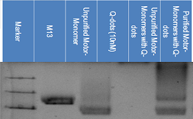
<p class="caption">Fig.2-1-3. | <p class="caption">Fig.2-3-3. 1% agarose gel electrophoresis of Motor-Monomers and Q-dots(Cy5)</p> | ||
</div> | </div><br> | ||
<div class="centerbox"> | |||
<p class="image"><img src="http://openwetware.org/images/e/e2/Fig.2-3-4.png" width=“280px” height="173px"></p> | |||
<p class="caption">Fig.2-3-4. 1% agarose gel electrophoresis of Motor-Monomers and Q-dots(EtBr)</p> | |||
</div><br> | |||
<div class="centerbox"> | <div class="centerbox"> | ||
<p class="image"><img src=""></p> | <p class="image"><img src="http://openwetware.org/images/6/6e/Fig.2-3-5.png" width=“280px” height="173px"></p> | ||
<p class="caption">Fig.2- | <p class="caption">Fig.2-3-5. 1% agarose gel electrophoresis of Motor-Monomers and Q-dots(EtBr)</p> | ||
</div> | </div><br> | ||
<br clear="right"> | <br clear="right"> | ||
<p class="paragraph"> | <p class="paragraph"> | ||
The first picture shows the existence of Qdots and the second picture shows the existence of DNA. Comparing the fourth, fifth and sixth lane from the left in the first picture, we can see the difference in the position of the Qdots. Comparing the fifth and sixth lane from the left in the two pictures, we can see the position of DNA, in this case, Motor-Monomers, and the Q-dots in the same position. This data shows that the Qdots connected to the Motor-Monomers through the connection of biotin and streptavidin. | The first picture shows the existence of Qdots and the second picture shows the existence of DNA. Comparing the fourth, fifth and sixth lane from the left in the first picture, we can see the difference in the position of the Qdots. Comparing the fifth and sixth lane from the left in the two pictures, we can see the position of DNA, in this case, Motor-Monomers, and the Q-dots in the same position. This data shows that the Qdots connected to the Motor-Monomers through the connection of biotin and streptavidin. | ||
</p> | </p> | ||
<br><br><br><br><br><br><br> | |||
<br> | |||
<br> | |||
< | |||
< | |||
<br> | |||
<h1 class="sub">2-4. Polymerization in solution</h1> | <h1 class="sub">2-4. Polymerization in solution</h1> | ||
| Line 357: | Line 367: | ||
<div class="centerbox"> | <div class="centerbox"> | ||
<p class="image"><img src=""></p> | <p class="image"><img src="http://openwetware.org/images/e/e1/Ex2-4-1.png"></p> | ||
<p class="caption">Fig.2-4-1. Agarose gel stained with EtBr shows the dimer band.</p> | <p class="caption">Fig.2-4-1. Agarose gel stained with EtBr shows the dimer band.</p> | ||
</div> | </div><br> | ||
<div class="centerbox"> | <div class="centerbox"> | ||
<p class="image"><img src="http://openwetware.org/images/4/45/2-4-2.png"></p> | <p class="image"><img src="http://openwetware.org/images/4/45/2-4-2.png"></p> | ||
<p class="caption">Fig.2-4-2. Ratio of the Monomer and the Dimer.</p> | <p class="caption">Fig.2-4-2. Ratio of the Monomer and the Dimer.</p> | ||
</div> | </div><br> | ||
<div class="centerbox"> | <div class="centerbox"> | ||
<p class="image"><img src=""></p> | <p class="image"><img src="http://openwetware.org/images/9/90/2-4-3.png"></p> | ||
<p class="caption">Fig.2-4-3. Relationship between polymerization degree and ratio of Polymer.</p> | <p class="caption">Fig.2-4-3. Relationship between polymerization degree and ratio of Polymer.</p> | ||
</div> | </div><br> | ||
<div class="centerbox"> | <div class="centerbox"> | ||
<p class="image"><img src=""></p> | <p class="image"><img src="http://openwetware.org/images/d/d4/2-4-4.png" width=“300px” height="200px"></p> | ||
<br><br> | |||
<p class="caption">Fig.2-4-4. TEM image shows tetramer of the Motor-Monomers.</p> | <p class="caption">Fig.2-4-4. TEM image shows tetramer of the Motor-Monomers.</p> | ||
</div> | </div> | ||
<div class="centerbox"> | <div class="centerbox"> | ||
<p class="image"><img src=""></p> | <p class="image"><img src="http://openwetware.org/images/4/4a/2-4-5.png" width=“300px” height="200px"></p> | ||
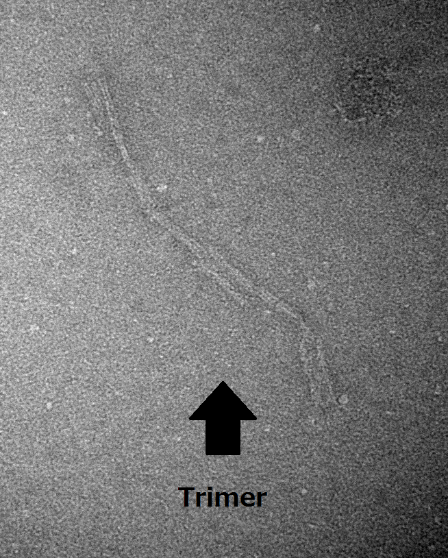
<p class="caption">Fig.2-4-5. TEM image shows trimer of the Motor-Monomers.</p> | <br><br> | ||
<p class="caption">Fig.2-4-5. TEM image shows trimer of the Motor-Monomers.</p> | |||
<p class="paragraph"> | </div> | ||
<br> | |||
<br><p class="paragraph"> | |||
Figure 2-4-1 and fig.2-4-2 show that the Motor-Monomer polymerized into the Motor-Polymer when streptavidin were added into Motor-Monomer solution. | Figure 2-4-1 and fig.2-4-2 show that the Motor-Monomer polymerized into the Motor-Polymer when streptavidin were added into Motor-Monomer solution. | ||
TEM analysis suggest that the Motor-Monomer without streptavidin were not polymerized in solution, while polymerized upon SA addition, trimer and tetramer were observed in SA(+) sample.</p> | TEM analysis suggest that the Motor-Monomer without streptavidin were not polymerized in solution, while polymerized upon SA addition, trimer and tetramer were observed in SA(+) sample.</p> | ||
| Line 386: | Line 397: | ||
<div align="center"> | |||
<a href="http://openwetware.org/wiki/Biomod/2014/Kashiwa/Receptor"><img src="http://openwetware.org/images/0/0c/ReceptorswitchKashiwa.png" onmouseover="this.src='http://openwetware.org/images/a/a3/Receptorswitch2Kashiwa.png'" onmouseout="this.src='http://openwetware.org/images/0/0c/ReceptorswitchKashiwa.png'" height="160px" width="240px" style="padding:5px 60px;"></a> | |||
</div> | |||
</body> | </body> | ||
Latest revision as of 16:22, 26 October 2014
<html> <head> <div id="#top"></div>
<script type="text/javascript" src="http://code.jquery.com/jquery-1.9.1.min.js"></script>
<script type="text/javascript"> new Image(120,80) = "http://openwetware.org/images/7/7a/Logo2Kashiwa.png";
new image(120,80) = "http://openwetware.org/images/1/1d/Logo2.5.png";
new image(71,143) = "http://openwetware.org/images/2/2a/Top1.gif";
new image(71,143) = "http://openwetware.org/images/f/f0/Top2.gif";
new image() = "http://openwetware.org/images/3/38/BackgroundKashiwa.png";
new image() = "http://openwetware.org/images/9/9c/BackgroundKashiwa2.png"; </script>
<script type="text/javascript">
  $(function(){
     $("#menu li").hover(function(){
        $("ul",this).show();
     },
     function(){
        $("ul",this).hide();
     });
  });
</script>
<script type="text/javascript">
<!--
if (document.images) {
// 設定開始(使用する画像を設定してください)
// 通常の画像 var img0 = new Image(120,80); img0.src = "http://openwetware.org/images/e/ec/LogoKashiwa.png";
// ポイント時の画像1 var img1 = new Image(120,80); img1.src = "http://openwetware.org/images/2/26/Logo3Kashiwa.png";
// 設定終了
} // ポイント時の処理 function On(name) { if (document.images) { document.images['def'].src = eval(name + '.src'); } } // 放した時の処理 function Off() { if (document.images) { document.images['def'].src = img0.src; } } // --> </script>
<META http-equiv="Content-Style-Type" content="text/css">
<style type="text/css">
.page-back {
position: fixed;
bottom: 25px;
right: 25px;}
p.paragraph {
 font-size :100%;
 line-height: 135%;
 margin: 10px 25px;
}
p.reference {
 font-size :90%;
 line-height: 135%;
 margin: 10px 20px;
}
p.indent-paragraph {
 font-size :110%;
 line-height:1.5;
 margin:0 30px;
 text-indent: 1em;
 }
h1.title a{
 font-size :100%;
 display: block;
 text-decoration: none;
 color: #000000;
font-weight:bolder;
 border-left: solid 5px #e00000;
 }
h2.reference a{
 font-size :90%;
 display: block;
 text-decoration: none;
 border-left: solid 5px;
 }
body {
 font-size: 12px;
 font-family: Frutiger, Helvetica, Arial;
 background-color: #B5D3FF;
 overflow-y:scroll;
 overflow-x:hidden;
} article {
background-color: #ffffff
} .container {
  background-color: #ffffff;
  margin-top:0px
}
.OWWNBcpCurrentDateFilled {display: none;}
}
- column-content
{
  width: 0px;
  margin: 0;
  padding: 0;
  float:left;
} .firstHeading {
  display:none;
  width:0px;
}
- globalWrapper
{
  width:900px;
  background-color: #ffffff;
  margin-left: auto;
  margin-right: auto;
}
- column-one
{
  display:none;
  width:0px;
  background-color: #ffffff;
}
- content
{
  margin: 0px;
  align: center;
  padding: 0px 12px;
  width:876px;
  border: 0;
}
- bodyContent
{
  width: 800px;
  padding: 0px 38px;
  align: center;
  background-color: #ffffff;
  position:relative;
}
- column-content
{
  width: 900px;
  background-color: #ffffff;
}
- footer
{
  position: center;
  width: 900px;
}
div.menubar {
  position: fixed;
  background-color: none; /* バーの背景色 */
  opacity: 0.95;
  border-top: 0px double white; /* バーの上端線 */
  border-bottom: 0px double white; /* バーの下端線 */
  min-width: 900px; /* メインメニュー全部が収まる最低横幅 */
  z-index:3
}
div.menubar ul#menu {
  margin: -33px 0px 0px -50px; /* メニューバー外側の余白 */
  padding: 0px; /* メニューバー内側の余白 */
  height: 80px; /* メニューバーの高さ */
  list-style-type: none;
}
div.menubar ul#menu li {
  min-width: 110px; /* メニュー項目の横幅 */
  height: 80px; /* メニュー項目の高さ(「メニューバーの高さ」と一致させる) */
  float: left;
  list-style-type: none;
  list-style-image: none;
  position: relative;
}
div.menubar ul#menu a {
  text-decoration: none; /* メニュー項目の装飾(下線を消す) */
  display: block;
  background-color: #1F003E;/* メニュー項目の背景色 */
  color: white; /* メニュー項目の文字色 */
  line-height: 80px; /* メニュー項目のリンクの高さ(「メニュー項目の高さ」と一致させる) */
  text-align: center; /* メインメニューの文字列の配置(中央寄せ) */
  width: 100%;
  height: 100%;
}
div.menubar ul#menu a:hover {
  background-image: url("http://openwetware.org/images/3/38/BackgroundKashiwa.png"); /* メニュー項目にマウスが載ったときの背景色 */
  color: #8B008B; /* メニュー項目にマウスが載ったときの文字色 */
}
div.menubar ul#menu a:active {
  background-image: url("http://openwetware.org/images/3/33/Background2.png"); /* メニュー項目にマウスが載ったときの背景色 */
  color: #8B008B; /* メニュー項目をクリックした時の文字色 */
}
/* メニューバー直後のClearfix */ div.menubar ul#menu {
zoom:1;
} div.menubar ul#menu:after {
height: 0; visibility: hidden; content: "."; display: block; clear: both;
}
div.menubar ul#menu ul.sub {
  background-color: #1F003E; /* サブメニュー全体の背景色 */
  border-width: 0px 0px 0px 0px; /* サブメニュー全体の枠線の太さ */
  border-style: solid; /* サブメニュー全体の枠線の線種 */
  border-color: #191970; /* サブメニュー全体の枠線の色 */
  margin: 0px;
  padding: 0px;
  display: none;
  position: absolute;
}
div.menubar ul#menu ul.sub li {
  width: 110px;
  height: 50px; /* サブメニュー1項目の高さ */
  border-width: 0px 0px 0px 0px; /* サブメニュー1項目の枠線の太さ */
  border-style: solid; /* サブメニュー1項目の枠線の線種 */
  border-color: #191970; /* サブメニュー1項目の枠線の色 */
  z-index: 3
}
div.menubar ul#menu ul.sub li a {
  line-height: 50px; /* サブメニュー1項目の行の高さ(「サブメニュー1項目の高さ」と合わせる) */
  text-align: center;  /* サブメニュー1項目の項目名の配置(中央寄せ) */
  text-indent: 0px;  /* サブメニュー1項目の項目名前方の余白 */
}
div.menubar ul#menu ul.sub li a:hover {
  background-image: ('back');
  background-size: 100% auto;
  background-color: #FF00FF; /* サブメニュー項目にマウスが載ったときの背景色 */
  color:  #8B008B; /* サブメニュー項目にマウスが載ったときの文字色 */
}
div.menubar ul#menu ul.sub li a:active {
  background-image: ('backclick') /* メニュー項目にマウスが載ったときの背景色 */
  backround-size: 100% auto;
  color: #8B008B; /* メニュー項目をクリックした時の文字色 */
}
</style>
<div class="menubar">
<ul id="menu">
<font face="Frutiger,Helvetica,Arial">
<li><a href="http://openwetware.org/wiki/Biomod/2014/Kashiwa"><img src="http://openwetware.org/images/e/ec/LogoKashiwa.png" onmouseover="this.src='http://openwetware.org/images/7/7a/Logo2Kashiwa.png'" onclick="this.src='http://openwetware.org/images/1/1d/Logo2.5.png'" onmouseout="this.src='http://openwetware.org/images/e/ec/LogoKashiwa.png'" height="80px" width="120px" name="def"></a>
</li>
<li><a href="http://openwetware.org/wiki/Biomod/2014/Kashiwa/Project" onMouseOver="On('img1')" onMouseOut="Off()"><span style="font-size:12pt;">PROJECT</span></a>
<ul class="sub">
<li><a href="http://openwetware.org/wiki/Biomod/2014/Kashiwa/Project#1" onMouseOver="On('img1')" onMouseOut="Off()"><span style="font-size:12pt;">Background</span></a></li>
<li><a href="http://openwetware.org/wiki/Biomod/2014/Kashiwa/Project#2" onMouseOver="On('img1')" onMouseOut="Off()"><span style="font-size:12pt;">Motivation</span></a></li>
<li><a href="http://openwetware.org/wiki/Biomod/2014/Kashiwa/Project#3" onMouseOver="On('img1')" onMouseOut="Off()"><span style="font-size:12pt;">Project Goals</span></a></li>
</ul>
</li>
<li><a href="http://openwetware.org/wiki/Biomod/2014/Kashiwa/Trial" onMouseOver="On('img1')" onMouseOut="Off()"><span style="font-size:12pt;"> EARLY TRIAL </span></a>
<ul class="sub">
<li><a href="http://openwetware.org/wiki/Biomod/2014/Kashiwa/Trial#1" onMouseOver="On('img1')" onMouseOut="Off()"><span style="font-size:12pt;">Design</span></a></li>
<li><a href="http://openwetware.org/wiki/Biomod/2014/Kashiwa/Trial#2" onMouseOver="On('img1')" onMouseOut="Off()"><span style="font-size:12pt;">Approaches</span></a></li>
</ul>
</li>
<li><a href="http://openwetware.org/wiki/Biomod/2014/Kashiwa/Design" onMouseOver="On('img1')" onMouseOut="Off()"><span style="font-size:12pt;">DESIGN</span></a>
<ul class="sub">
<li><a href="http://openwetware.org/wiki/Biomod/2014/Kashiwa/Design#2" onMouseOver="On('img1')" onMouseOut="Off()"><span style="font-size:12pt;">The Receptor</span></a></li>
<li><a href="http://openwetware.org/wiki/Biomod/2014/Kashiwa/Design#1" onMouseOver="On('img1')" onMouseOut="Off()"><span style="font-size:12pt;">The Motor</span></a></li>
</ul>
</li>
<li><a href="http://openwetware.org/wiki/Biomod/2014/Kashiwa/Highlights" onMouseOver="On('img1')" onMouseOut="Off()"><span style="font-size:12pt;"> EXPERIMENT </span></a>
<ul class="sub">
<li><a href="http://openwetware.org/wiki/Biomod/2014/Kashiwa/Highlights" onMouseOver="On('img1')" onMouseOut="Off()"><span style="font-size:12pt;">Highlights</span></a></li>
<li><a href="http://openwetware.org/wiki/Biomod/2014/Kashiwa/Receptor" onMouseOver="On('img1')" onMouseOut="Off()"><span style="font-size:12pt;">The Receptor</span></a></li>
<li><a href="http://openwetware.org/wiki/Biomod/2014/Kashiwa/Motor" onMouseOver="On('img1')" onMouseOut="Off()"><span style="font-size:12pt;">The Motor</span></a></li>
</ul>
<li><a href="http://openwetware.org/wiki/Biomod/2014/Kashiwa/Discussion" onMouseOver="On('img1')" onMouseOut="Off()"><span style="font-size:12pt;"> DISCUSSION </span></a>
<ul class="sub">
<li><a href="http://openwetware.org/wiki/Biomod/2014/Kashiwa/Discussion#1" onMouseOver="On('img1')" onMouseOut="Off()"><span style="font-size:12pt;">Achievements</span></a></li>
<li><a href="http://openwetware.org/wiki/Biomod/2014/Kashiwa/Discussion#2" onMouseOver="On('img1')" onMouseOut="Off()"><span style="font-size:12pt;">Future</span></a></li>
</ul>
</li>
<li><a href="http://openwetware.org/wiki/Biomod/2014/Kashiwa/Protocols" onMouseOver="On('img1')" onMouseOut="Off()"><span style="font-size:12pt;">PROTOCOL</span></a>
</li>
<li><a href="http://openwetware.org/wiki/Biomod/2014/Kashiwa/Team" onMouseOver="On('img1')" onMouseOut="Off()"><span style="font-size:12pt;">TEAM</span></a>
<ul class="sub">
<li><a href="http://openwetware.org/wiki/Biomod/2014/Kashiwa/Team#1" onMouseOver="On('img1')" onMouseOut="Off()"><span style="font-size:12pt;">Members</span></a></li>
<li><a href="http://openwetware.org/wiki/Biomod/2014/Kashiwa/Team#2" onMouseOver="On('img1')" onMouseOut="Off()"><span style="font-size:12pt;">Sponsors</span></a></li>
</ul>
</li>
</font>
</ul>
</div> <br> <br> <br> <div class="page-back"><a href="#top"> <img src="http://openwetware.org/images/2/2a/Top1.gif" onmouseover="this.src='http://openwetware.org/images/f/f0/Top2.gif'" onmouseout="this.src='http://openwetware.org/images/2/2a/Top1.gif'" height="150px"/></a></div> </head>
</html>
<html>
<head>- <style>
div.imagebox {
  border: 1.7px dashed #2E2EFE; /* 1.枠線 */
  background-color: #F0F8FF;  /* 2.背景色 */
  width: 290px; /* 横幅 */
  float: right; /* 右に配置 */
  margin: 0px 30px 0px 10px;
}
div.centerbox {
  border: 1.7px dashed #2E2EFE; /* 1.枠線 */
  background-color: #F0F8FF;  /* 2.背景色 */
  width: 290px; /* 横幅 */
  float: left; /* 右に配置 */
  margin: 0px 50px;
}
div.autobox{
  border: 1.7px dashed #2E2EFE; /* 1.枠線 */
  background-color: #F0F8FF;  /* 2.背景色 */
  width: 300px; /* 横幅 */
  height: 300px;
  float: right; /* 右に配置 */
  margin: 0px 30px 0px 10px;
}
p.image {
  text-align: center;
  width: 280px;
  height: 173px;
  margin: 5px;
}
p.caption {
  margin: 5px 10px;
  font-size: 90%;  /* 5.文字サイズ */
  color: black; /* 6.文字色 */
}
p.cap {
font-size: 90%;
}
h1.big{
 font-size :16px;
 text-decoration: none;
 color: #000000;
 margin: 10px 15px;
 font-weight: bolder;
 }
h1.sub {
 font-size:20px;
 font-weight: bolder;
 margin: 10px 5px;
}
p.menu {
 font-size :100%;
 line-height: 135%;
 margin: 0px 40px;
}
</style> </head>
<body>
EXPERIMENTS
<a name="background"> The Moving System: The Motor</a>
<img src="http://openwetware.org/images/5/57/Monomerexperiments.png" width="300px" height="170px" align="right" style="margin:10px;">
This section describes the details of our approach to develop the moving system "Motor" of PoLICe. The approach mainly consists of three steps as follows.
- <a href="#2-1">2-1. Production of the Motor-Monomer </a>
- <a href="#2-1-1"> 2-1(a). Folding of the Motor-Monomer body </a>
- <a href="#2-1-2"> 2-1(b). Synthesis of divalent SA </a>
- <a href="#2-1-3"> 2-1(c). Equipment of the Motor-Monomer with divalent SA </a>
<a name="#2-1"></a><a name="2-1"></a>
- <a href="#2-2">2-2. Deactivation and reactivation of the binding activity of streptavidin</a>
- <a href="#2-3">2-3. Incorporation of the Motor-Monomers into the liposome </a> <a name="2-1-1"></a>
2-1. Production of the Motor-Monomer
Producing the Motor-Monomer is divided into three sections: Folding the Motor-Monomer body, synthesizing divalent streptavidin and equipping the Motor-Monomer body with divalent streptavidin.
2-1(a). Folding of the Motor-Monomer body
| <img src="http://openwetware.org/images/2/25/Motorkashiwa1.png" width="180px"> | <img src="http://openwetware.org/images/d/d9/Motorkashiwa2.png" width="180px"> | <img src="http://openwetware.org/images/e/e8/Motorkashiwa3.png" width="180px"> | <img src="http://openwetware.org/images/e/ef/Motorkashiwa4.png" width="180px"> |
|---|---|---|---|
Fig.2-1(a)-1. Gel analysis of the Monomers annealed in different time. | Fig.2-1(a)-2. Gel analysis of the Monomers annealed in different temperature. | Fig.2-1(a)-3. Gel analysis of the Monomers annealed in different concentration of NaCl. | Fig.2-1(a)-4. Gel analysis of the Monomers annealed in different concentration of MgCl2. |
In this experiment, the assembly condition of the Motor-Monomer structure was optimized and results were analyzed by agarose gel electrophoresis. The optimum conditions were confirmed by comparing migration distances of each samples. The sample of which migration distance is the longest was regarded as the optimum condition.
| <IMG src="http://openwetware.org/images/c/ce/Monomer_temforwiki.JPG" widh="300"/> |
Fig.2-1(a)-5. TEM image for the Motor-Monomer. |
The optimum results are as follows.
- Concentration of MgCl2 : 15 mM
- Temperature of annealing : 49.1 °C
- Time for annealing : 2 hours.
- Concentration of NaCl : 2.5 mM.
<a name="2-1-2"></a> The folding is corroborated by the TEM image (in fig.2-1(a)-5.)
2-1(b). Synthesis of divalent streptavidin
<img src="http://openwetware.org/images/b/ba/2-1%28b%29-1.png">
Fig.2-1(b)-1. SDS-PAGE showing purified divalent SA.
In this experiment, divalent streptavidin (SA) was synthesized for equipment to the Motor-Monomers.
TaKaRa Competent Cell BL21 was used for protein expression. The purification of divalent SA on the nickel-affinity column was analyzed by SDS-PAGE.
<img src="http://openwetware.org/images/2/28/2-1%28b%29-2.png">
Fig.2-1(b)-2. SDS-PAGE showing the affinity of divalent SA with biotin-modified oligos.
The affinity of divalent SA with biotin-modified oligonucleotides was then analyzed by SDS-PAGE.
<a name="2-1-3"></a>
2-1(c). Equipment of the Motor-Monomer with SA
We used click reaction, alkyne-azide huisgen cycloaddition, to equip the Motor-Monomer with SA. The click reaction was evaluated by the following steps:
(i) Optimization of condition for click reaction
<img src="http://openwetware.org/images/7/72/Click1.png" width="700px">
Fig.2-1(c)-1. Optimum combinations of click reaction.
First, the optimum combination of click reaction between simple alkyne and azide was analyzed. two pairs out of 16 pairs were consequently chosen as the optimum combinations.
<img src="http://openwetware.org/images/a/a7/2-1%28c%2922.png">
Fig.2-1(c)-2. Analysis of click reaction by Native-PAGE (16 pairs.)
<img src="http://openwetware.org/images/1/16/2-1%28c%2932.png">
Fig.2-1(c)-3. Analysis of click reaction by Native-PAGE (4 pairs.)
<img src="http://openwetware.org/images/f/f9/2-1%28c%2942.png">
Fig.2-1(c)-4. Analysis of click reaction by Native-PAGE (2 pairs.)
<img src="http://openwetware.org/images/5/55/Table2-1-c-1.png" width=“160px” height="100px">
Table 2-1(c)-1
<img src="http://openwetware.org/images/e/ea/Gonza_click.png">
Fig.2-1(c)-5.Relation between time and rate of reaction
In this experiment, as the reaction was regarded as pseudo-first-order reaction, trendline was fitted and kon was gotten. There were few difference between 2pairs in reactivity.
(ii) Evaluation of biotin-binding activity of divalent SA
<img src="http://openwetware.org/images/8/8b/2-1%28c%2962.png">
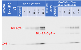
Fig.2-1(c)-6. Analysis of NHS reaction by Native-PAGE (Cy5).
<img src="http://openwetware.org/images/c/c8/2-1%28c%2972.png">
Fig.2-1(c)-7. Analysis of NHS reaction by Native-PAGE (SYBR Gold).
Figure 2-1(c)-1 shows that SA was linked with Cy5-modified oligonucleotide via click reaction. In Fig.2-1(c)-2, because the lane of only Cy5 and NH2-modified oligonucleotide is not stained, the stained bands is from biotin-oligonucleotide. This picture shows modified SA can bind biotin. It may show the activity to bind biotin decreases as the amount of modification of SA increases, but the experiment of modification SA with Cy5-NHS deny. The reason of that may be derived from steric hindrance of Cy5-oligonucleotide which binds to SA.
(iii) Analysis of click chemistry of alkyne and azide-modified Motor-Monomer
Click reaction between azide attached to Motor-Monomer and alkyne was confirmed as a preliminary step of modification of Motor-Monomer with inactivated SA.
<img src="http://openwetware.org/images/3/33/Gonzaclick2.png">
Fig.2-1(c)-8. Agarose gel labelled with Cy5.
<img src="http://openwetware.org/images/9/90/Gonzaclick.png">
Fig.2-1(c)-9. Agarose gel stained with EtBr.
The figure shows that the band of Cy5 and Motor-Monomer in lane of 30 h are in the same place. It means that alkyne-Cy5 oligo was attached to Motor-Monomer by click reaction.
(iv) Analysis of click chemistry of azide and alkyne-modified deactivated SA
<img src="http://openwetware.org/images/4/49/2-1%28c%29-10.png">
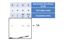
Fig.2-1(c)-10. Agarose gel labeled with Cy3.
<img src="http://openwetware.org/images/5/58/2-1%28c%29-11.png">
Fig.2-1(c)-11. Agarose gel labeled with Cy5.
<img src="http://openwetware.org/images/0/0d/2-1%28c%29-12.png">
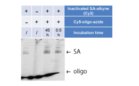
Fig.2-1(c)-12. Agarose gel labeled with EtBr.
The gel images show that the band of Cy3 (inactivated SA) and Cy5 (azide) appeared at the same position . And FRET was observed in the lane of 45 h incubation. These results indicates that Cy5-azide was attached to inactivated SA by click chemistry.
<a name="2-2"></a>It therefore suggests that alkyne-modified deactivated SA can react with Cy5-azide. And rate of reaction reaches 60 % after 45 hours incubation.
These figures confirm the activity of modified SA to bind biotin-nucleotide.
2-2. Deactivation and activation of the binding activity of streptavidin
<img src="http://openwetware.org/images/8/87/Inactivated-SA-name.png">
Fig.2-2-1. Mechanism of this experiment.
In this experiment, deactivation and reactivation of streptavidin (SA) were confirmed by Native PAGE.
First, two complementary oligonucleotides having biotin or desthiobiotin were annealed. The oligos having biotin is named Linker and the oligos having desthiobiotin is named Blocker. The resulting dsDNA was then mixed with divalent SA for deactivation. After removing excess SA with biotin-beads, ds-DNA bound to divalent SA was cut by HindⅢ for reactivation. Another oligonucleotides having biotin, named Chaser, were then added to replace Blocker.
Blocker was labeled with Cy3 and Chaser was labeled with Cy5 to confirm the replacement by Native-PAGE.
| <img src="http://openwetware.org/images/4/48/Inactivated-SA-cy3.png"> | <img src="http://openwetware.org/images/e/e1/Inactivated-SA-cy5.png"> |
|---|---|
| Fig.2-2-2. Cy3 fluorescence image of Native-PAGE. | Fig.2-2-3. Cy5 fluorescence image of Native-PAGE. |
In fig.2-2-3, multiple bands of SA-dsDNA complexes appears in lane 3 which is caused by non-ideal complexes. It is considered that biotin-beads successfully distinguished the ideal SA-dsDNA complex because the upper band in lane 3 mostly disappeared in lane 4.
In fig.2-2-4, a band at the same position of the ideal SA-dsDNA complex is only shown in lane 7. We therefore confirmed the<a name="2-3"></a> replacement of Blocker with Chaser after cutting dsDNA by HindⅢ and that the replacement did not happen without HindⅢ.
2-3. Incorporation of the Motor-Monomers into the liposome
In this experiment, Motor-Monomers which have biotin staple combined with Q-dot connected with streptavidin were put into the giant unilamellar vesicles (GUV). Inclusion of Motor-Monomers was observed by confocal microscope.
GUVs were dyed by Nile Red. Yellow green dots indicate the fluorescence of Nile Red and red dots indicate the fluorescence of Q-dots combined with Motor-Monomers.
<img src="http://openwetware.org/images/f/f1/Fig.2-3-1.png">
Fig.2-3-1. Confocal microscope image of GUV including Motor-Monomers.
<img src="http://openwetware.org/images/0/07/Fig.2-3-2.png">
Fig.2-3-2. Confocal microscope image of GUV not including Motor-Monomers.
Comparing the two pictures, we can see the red dots inside the GUV only in the picture showing the GUVs containing Motor-Monomers in the inner solution. From this fact, we could confirm the inclusion of Motor-Monomers into the GUV. As the excitation and fluorescence wave length of Nile Red and Qdots were close, the membrane of GUV was shown in red even in the picture of GUV not including the Q-dots.
The connection of Qdot to the Motor-Monomer was then confirmed by the agarose gel electrophoresis.
<img src="http://openwetware.org/images/b/b3/Fig.2-3-3.png" width=“280px” height="173px">
Fig.2-3-3. 1% agarose gel electrophoresis of Motor-Monomers and Q-dots(Cy5)
<img src="http://openwetware.org/images/e/e2/Fig.2-3-4.png" width=“280px” height="173px">
Fig.2-3-4. 1% agarose gel electrophoresis of Motor-Monomers and Q-dots(EtBr)
<img src="http://openwetware.org/images/6/6e/Fig.2-3-5.png" width=“280px” height="173px">
Fig.2-3-5. 1% agarose gel electrophoresis of Motor-Monomers and Q-dots(EtBr)
The first picture shows the existence of Qdots and the second picture shows the existence of DNA. Comparing the fourth, fifth and sixth lane from the left in the first picture, we can see the difference in the position of the Qdots. Comparing the fifth and sixth lane from the left in the two pictures, we can see the position of DNA, in this case, Motor-Monomers, and the Q-dots in the same position. This data shows that the Qdots connected to the Motor-Monomers through the connection of biotin and streptavidin.
2-4. Polymerization in solution
Polymerization of the Motor-Monomers to the Motor-Polymer was confirmed as a preliminary step of polymerization in a liposome.
<img src="http://openwetware.org/images/e/e1/Ex2-4-1.png">
Fig.2-4-1. Agarose gel stained with EtBr shows the dimer band.
<img src="http://openwetware.org/images/4/45/2-4-2.png">
Fig.2-4-2. Ratio of the Monomer and the Dimer.
<img src="http://openwetware.org/images/9/90/2-4-3.png">
Fig.2-4-3. Relationship between polymerization degree and ratio of Polymer.
<img src="http://openwetware.org/images/d/d4/2-4-4.png" width=“300px” height="200px">
Fig.2-4-4. TEM image shows tetramer of the Motor-Monomers.
<img src="http://openwetware.org/images/4/4a/2-4-5.png" width=“300px” height="200px">
Fig.2-4-5. TEM image shows trimer of the Motor-Monomers.
Figure 2-4-1 and fig.2-4-2 show that the Motor-Monomer polymerized into the Motor-Polymer when streptavidin were added into Motor-Monomer solution. TEM analysis suggest that the Motor-Monomer without streptavidin were not polymerized in solution, while polymerized upon SA addition, trimer and tetramer were observed in SA(+) sample.
The results suggests that the polymerization were proceeded in solution by adding divalent SA.
<a href="http://openwetware.org/wiki/Biomod/2014/Kashiwa/Receptor"><img src="http://openwetware.org/images/0/0c/ReceptorswitchKashiwa.png" onmouseover="this.src='http://openwetware.org/images/a/a3/Receptorswitch2Kashiwa.png'" onmouseout="this.src='http://openwetware.org/images/0/0c/ReceptorswitchKashiwa.png'" height="160px" width="240px" style="padding:5px 60px;"></a>
</body>
<footer style="position:relative; left:600px;"> © 2014 UTokyo Chem & Bio </footer>