Beauchamp:Lab Members: Difference between revisions
No edit summary |
Sarah Baum (talk | contribs) |
||
| (37 intermediate revisions by 2 users not shown) | |||
| Line 4: | Line 4: | ||
|-valign="top" | |-valign="top" | ||
|width=179.25px style="padding: 5px; background-color: #ffffff; border: 2px solid #CC6600;" | | |width=179.25px style="padding: 5px; background-color: #ffffff; border: 2px solid #CC6600;" | | ||
<h3>Lab Photos</h3> | <h3>Current Lab Photos</h3> | ||
#[http://picasaweb.google.com/BeauchampLab Click Here] | #[http://picasaweb.google.com/BeauchampLab Click Here] | ||
<h3>Current Lab Members</h3> | |||
#[http://nba.uth.tmc.edu/resources/faculty/members/beauchamp.htm Web Page for Michael Beauchamp hosted by the University of Texas Department of Neurobiology and Anatomy] | |||
#[http://www.openwetware.org/wiki/Image:Beauchamp_Dec2011.pdf Click Here to download Michael Beauchamp's curriculum vitae] | |||
#[[User:Sarah Baum|Sarah Baum]] | |||
#Inga Schepers | |||
#John Magnotti | |||
<h3>Lab Alumni</h3> | |||
#[[User:Audrey Nath|Audrey Nath]] | |||
#[[User:Siavash Pasalar|Siavash Pasalar]] | |||
#[[User:Eswen Fava|Eswen Fava]] | |||
#[[User:Westley|J. Westley Ohman]] | |||
#[[User:Molly|Molly Bryan]] | |||
#[[User:Nafi|Nafi Yasar]] | |||
#Dona Murphey | |||
#Neel Kishan http://cns.bu.edu/~nkishan | |||
#Ashley Kingon | |||
==Lab Photos== | |||
[[Image: OHBMSeattle.jpg | 600px]] | |||
Sarah, Inga, and John at OHBM 2013 in Seattle. | |||
[[Image:NRC2012.jpg | 600 px]] | |||
Debshila, Josephine, Mike, Sarah, and Inga at the NRC Poster Competition 2012. Photo by Dwight C. Andrews/The University of Texas Medical School at Houston Office of Communications. | |||
[[Image:mcgurkmeme.jpg]] | |||
Hipster dog knows why you don't get the McGurk effect. | |||
[[Image:shootingtrip.jpg | 600 px]] | |||
[[Image: GreatWall.jpg | 600 px]] | |||
Mike and Sarah at the Great Wall during OHBM 2012. | |||
==Old Lab Photos== | |||
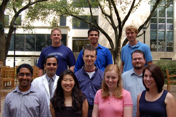
[[Image: Summerlab5.jpg | 600 px]] | |||
[ | [[Image:MaryMikeAndSuma.jpg| 600 px| "Cortex Interruptus"]] | ||
[[Image:Beauchamp_12.jpg| 600 px| "Cortex Interruptus"]] | |||
[[Image:Group1MSB.jpg| 600px]] | |||
[[Image:PhotoAerial.jpg |200 px| "Medical Center Aerial Shot"]] | |||
[[image:OutsideMSB.jpg|200 px| "Outside Patio"]] | |||
[[image:Lobby1.jpg | 200 px| "MSB Lobby"]] | |||
[[Image:TanVipTho.jpg| 200 px| ]] | |||
== Videos == | |||
Near Death Experiences: | |||
: | |||
[[Image:NearDeath1.jpg| 500 px]] | |||
: | [http://video.google.com/videoplay?docid=-3693743977857556383&hl=en Part 4 link] | ||
== Projects == | |||
The human brain is one of the most complex biological machines, containing billions of neurons and trillions of synapses. The Beauchamp Lab studies the living human brain using a variety of techniques. The main technique is blood-oxygen level dependent functional magnetic resonance imaging (BOLD fMRI). The primary research interest of the laboratory is multisensory integration. While vision is our most important sensory modality (taking up about a quarter of our brain’s real estate) other sensory modalities are also important. For instance, even if we cannot see our mobile phone blinking, we can hear its ring or feel its vibration in our pocket. These different modalities are encoded by our brain in very different ways. The auditory system is most concerned with the frequency (high or low-pitched sounds), as is the tactile system (slow stroking vs. quick vibrations). In contrast, our visual system is organized by the spatial location of stimuli--a stroke may damage our ability to see objects on the left side of the room but not the right side. Although each sensory modality is organized fundamentally differently, our brain must integrate the information provided by the different modalities in order to make decisions. For instance, is the phone ringing or not? Our research has shown that regions of superior temporal sulcus are especially important for this process of multisensory integration. In superior temporal sulcus, different sensory inputs converge into patches of cortex, allowing multisensory integration to occur. Because BOLD fMRI offers only an indirect measure of neuronal activity, it is important to combine fMRI with other measures of brain function. In particular, we combine fMRI with electrical and magnetic stimulation and recording of brain activity. Patients with intractable epilepsy are frequently implanted with subdural electrodes. While implanted with the electrodes, these patients may consent to participate in our experiments. fMRI is performed implantation to localize the functional areas under the electrode. We have demonstrated that stimulating an electrode located over a brain areas sensitive to color (as shown with fMRI and local field potential recording) produces the percept of a color that matched the color that evoked the strongest response, a blue-purple hue. This demonstrates a close correspondence between neuronal selectivity and perception. Using transcranial magnetic stimulation (TMS) in combination with fMRI allows us to explore the effect of interfering with activity in specific brain areas on perception in normal human subjects. | |||
Revision as of 13:30, 21 June 2013
Current Lab PhotosCurrent Lab Members
Lab Alumni
Lab PhotosSarah, Inga, and John at OHBM 2013 in Seattle. Debshila, Josephine, Mike, Sarah, and Inga at the NRC Poster Competition 2012. Photo by Dwight C. Andrews/The University of Texas Medical School at Houston Office of Communications. Hipster dog knows why you don't get the McGurk effect.
Mike and Sarah at the Great Wall during OHBM 2012. Old Lab PhotosVideosNear Death Experiences: ProjectsThe human brain is one of the most complex biological machines, containing billions of neurons and trillions of synapses. The Beauchamp Lab studies the living human brain using a variety of techniques. The main technique is blood-oxygen level dependent functional magnetic resonance imaging (BOLD fMRI). The primary research interest of the laboratory is multisensory integration. While vision is our most important sensory modality (taking up about a quarter of our brain’s real estate) other sensory modalities are also important. For instance, even if we cannot see our mobile phone blinking, we can hear its ring or feel its vibration in our pocket. These different modalities are encoded by our brain in very different ways. The auditory system is most concerned with the frequency (high or low-pitched sounds), as is the tactile system (slow stroking vs. quick vibrations). In contrast, our visual system is organized by the spatial location of stimuli--a stroke may damage our ability to see objects on the left side of the room but not the right side. Although each sensory modality is organized fundamentally differently, our brain must integrate the information provided by the different modalities in order to make decisions. For instance, is the phone ringing or not? Our research has shown that regions of superior temporal sulcus are especially important for this process of multisensory integration. In superior temporal sulcus, different sensory inputs converge into patches of cortex, allowing multisensory integration to occur. Because BOLD fMRI offers only an indirect measure of neuronal activity, it is important to combine fMRI with other measures of brain function. In particular, we combine fMRI with electrical and magnetic stimulation and recording of brain activity. Patients with intractable epilepsy are frequently implanted with subdural electrodes. While implanted with the electrodes, these patients may consent to participate in our experiments. fMRI is performed implantation to localize the functional areas under the electrode. We have demonstrated that stimulating an electrode located over a brain areas sensitive to color (as shown with fMRI and local field potential recording) produces the percept of a color that matched the color that evoked the strongest response, a blue-purple hue. This demonstrates a close correspondence between neuronal selectivity and perception. Using transcranial magnetic stimulation (TMS) in combination with fMRI allows us to explore the effect of interfering with activity in specific brain areas on perception in normal human subjects. |


