When we unplugged the LCD display from the circuit board, the LCD display for the machine was no longer functional.
When we unplugged the white wire that connects the circuit board to the main heating block,the LCD displayed a reading of -40 degrees on the machine.
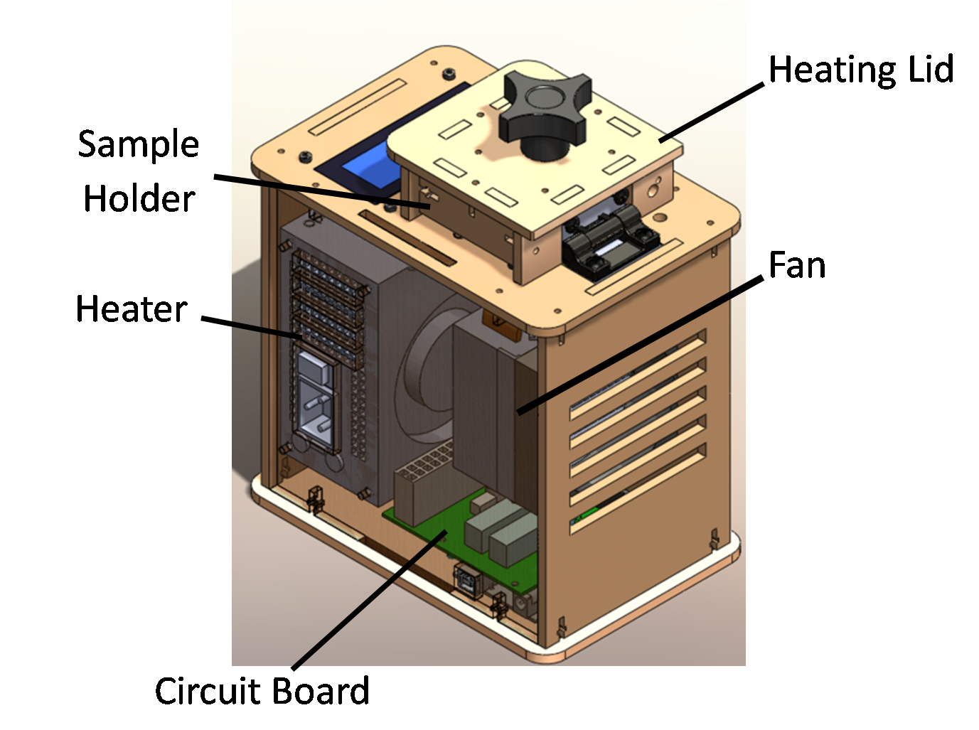
Test Run
(Write the date you first tested Open PCR and your experience(s) with the machine)
Protocols
Polymerase Chain Reaction
How it Works
In order to analyze genomes and genes,scientists have used polymerase chain reaction (PCR) techniques to amplify DNA. Multiple copies of DNA sequences much be made to be able to better view specific aspects of a gene due to the fact that isolated DNA samples are too small to view individually. In a test tube, PCR takes place by copying short fragments of DNA over and over again, also known as amplification. By altering the temperature within the test tubes, this allows the DNA strands to separate (boiling), bind to primers (cooled),and catalyze (warmed) to double the amount of DNA fragments after each cycle. By repeating each cycle the amount to DNA grows exponentially as two copies are made per strand.
How to Amplify a Patient's DNA
1. After the Patients DNA is placed in to the test tubes along with the Master Mix, the tubes are placed into the PCR machine.
2. As the machine warms to almost a boil, at 95 degrees Celsius, the DNA strands will begin to separate.
3. Once the PCR machine has cooled down to 57 degrees Celsius, the primer will bind to the target sequence.
4. The PCR machine needs to then warm back up to 72 degrees Celsius in order for extension of DNA to occur. This initiates the taq function where the taq protein, along with magnesium chloride, takes free floating nucleotides (DNTPs) and attaches them to the DNA stand in a reverse direction.
5. Once steps 2-4 are repeated several times, each cycle will create newly replicated DNA stands and allow replication to continue until Master Mix is used up.
PCR Master Mix Components
For a 50µl reaction volume:
-GoTaq® Colorless Master Mix: 25µl
-Upstream primer: 10µM
-Downstream primer: 10µM
-DNA template 1–5µl
-Nuclease-Free Water to 50µl
Substances in Test Tubes
Reagent
Volume
Template DNA (20 ng)
.2 μL
10 μM forward primer
1.0 μL
10 μM reverse primer
1.0 μL
GoTaq master mix
50.0 μL
dH20
47.8 μL
Total Volume
100.0 μL
Patient Numbers
Patient #29341 is female at the age of 53.
Patient #23292 is male at the age of 56.
Flourimeter
Flourimeter Setup
Flourmeter Assembly Procedure
(step by step)
Saving Images to ImageJ
1. Open ImageJ
2. Click file and then click open (and the files from your computer open up)
3. Click the desired picture and it will open up on ImageJ
Research and Development
Specific Cancer Marker Detection - The Underlying Technology
Polymerase Chain Reaction (PCR) works by adding primers, nucleotides, a catalyst, and a polymerase to a sample of DNA. Then the samples are placed in PCR machine which will cycle through the heats 95 degrees celsius to 57 degrees celsius to 72 degrees celsius and repeat that cycle thirty times. When the temperature is 95 degrees the DNA double helix denatures into individual single strands. After which, when the temperature is lowered to 57 degrees, the primers anneal to the single strand of DNA in preparation for replication. Then the sample is heated to 72 degrees celsius where TAQ polymerase copies all the DNA after the primers. Given everything else for the PCR reaction the success of reaction depends on whether or not the primers placed in the mixture match some segment on the DNA strand. In this case the primers match with the known r17879961 (CHEK2) cancer-related segment were placed into mixture with the DNA. Specifically the forward primer is: TGGTATAAGACATTCCTGTC and the reverse primer is: AACTCTTACACTGCATACAT. Therefore only DNA segments that match these primers will be amplified. This method of replicating DNA works by having the two primers anneal to different strands of DNA and replicating in one direction. This process leaves two partial strands of DNA which is not the target sequence, however, it does contain the target sequence. Over the next few cycles the other primer will anneal to the same strand of DNA (if the forward annealed to it first then the reverse will anneal to it and vice versa). After annealing it will copy in the other direction and end where the other primer started giving you the 200 base pair target sequence. Because only segments with the primers are amplified if the patient does not have this r17879961 single nucleotide polymorphism (SNP) the DNA will not be amplified. Therefore the positive result for having the r17879961 SNP is the presence of amplified DNA in the PCR reaction.
<http://openwetware.org/images/1/1f/Acetic_acid_db.jpeg>
(BONUS points: Use a program like Powerpoint, Word, Illustrator, Microsoft Paint, etc. to illustrate how primers bind to the cancer DNA template, and how Taq polymerases amplify the DNA. Screen-captures from the OpenPCR tutorial might be useful. Be sure to credit the source if you borrow images.)

